Molecular figures
Every figure in one place, to review before it goes into a topic. Each one is drawn from facts with textbook sources, and a mechanism steps through its true states.
3D model
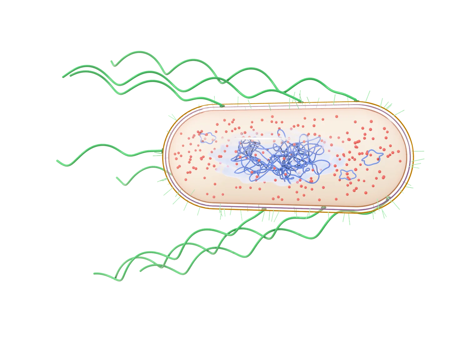
E. coli, a rod-shaped prokaryote, shown twice: as a model of the whole cell, which you can turn, and in a longitudinal section, a slice along its length. Its envelope has three layers: the outer membrane, a thin cell wall, and the plasma membrane. Inside, with no nucleus, a tangle of DNA, the nucleoid, fills the middle of the cytoplasm, with small rings of DNA, plasmids, and many ribosomes round it. Outside, short fimbriae cover the cell, and long wavy flagella rise all over it, each turned by a motor in the envelope, and stream back from it. Arrow keys turn it, plus and minus zoom, and Home brings back the first view.
Longitudinal section
Drag to turn it, pinch or scroll to zoom, and tap a part to read what it does.
E. coli, a prokaryote about 2 µm long, has no nucleus and no membrane-bound organelles: its one circular chromosome lies in the nucleoid, a region of the cytoplasm with no membrane round it. Its envelope is the plasma membrane, a thin wall of peptidoglycan and, outside that, the outer membrane of a gram-negative bacterium. Flagella, turned by motors in the envelope, drive it along, and fimbriae let it stick to surfaces and cells.
Simplified: not to scale: the envelope drawn thicker, and the flagella and fimbriae shorter than they are; the chromosome far shorter than it is: stretched out, it would be about 1.1 mm long, over 500 times the cell’s length; far fewer ribosomes than a real cell has; the flagella drawn apart, though while it swims they turn together in one bundle; in the model, the fimbriae shown only round its outline, where they stand out from it; the wall in carbohydrate’s colour, though its cross-links are peptides; the outer membrane drawn like the plasma membrane, though its outer layer holds lipopolysaccharide; ribosomes in RNA’s colour, though they are part protein.
10 sources
- Clark MA, Douglas M, Choi J. Biology 2e. OpenStax; 2018. 4.2 Prokaryotic Cells.
- Clark MA, Douglas M, Choi J. Biology 2e. OpenStax; 2018. 4.3 Eukaryotic Cells.
- Parker N, Schneegurt M, Thi Tu AH, et al. Microbiology. OpenStax; 2016. 3.3 Unique Characteristics of Prokaryotic Cells.
- Parker N, Schneegurt M, Thi Tu AH, et al. Microbiology. OpenStax; 2016. 3.4 Unique Characteristics of Eukaryotic Cells.
- Parker N, Schneegurt M, Thi Tu AH, et al. Microbiology. OpenStax; 2016. 14.3 Mechanisms of Antibacterial Drugs.
- Parker N, Schneegurt M, Thi Tu AH, et al. Microbiology. OpenStax; 2016. 9.2 Oxygen Requirements for Microbial Growth.
- Parker N, Schneegurt M, Thi Tu AH, et al. Microbiology. OpenStax; 2016. 15.2 How Pathogens Cause Disease.
- Clark MA, Douglas M, Choi J. Biology 2e. OpenStax; 2018. 7.4 Oxidative Phosphorylation.
- Clark MA, Douglas M, Choi J. Biology 2e. OpenStax; 2018. 14.2 DNA Structure and Sequencing.
- Escherichia coli. Wikipedia, citing Encyclopaedia Britannica and Kubitschek HE, J Bacteriol 1990.
3D model
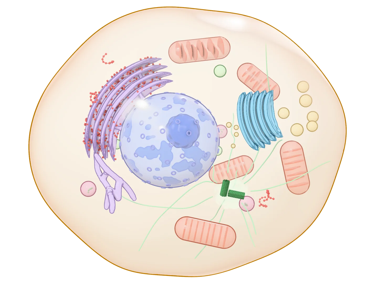
An animal cell, a eukaryote, shown twice: as a model of the whole cell, which you can turn, and in cross-section, as a slice through its middle. Its plasma membrane encloses the cytoplasm. The nucleus, inside a double envelope crossed by pores, holds the chromatin and a dense nucleolus. Beside it the rough endoplasmic reticulum lies in sheets studded with ribosomes, joined to the tubes of the smooth ER. The Golgi apparatus is a stack of curved sacs with vesicles round it, some carrying their load to the plasma membrane. Mitochondria show their folded inner membrane; lysosomes, peroxisomes and free ribosomes are scattered through the cytoplasm; and microtubules spread from the centrosome, with its two centrioles, by the nucleus. Arrow keys turn it, plus and minus zoom, and Home brings back the first view.
Cross-section
Drag to turn it, pinch or scroll to zoom, and tap a part to read what it does.
A eukaryotic cell, 10 to 100 µm across, keeps most of its DNA in a nucleus and shares its work among compartments bounded by membranes. Proteins for membranes and for export are made on the rough ER, modified in the Golgi apparatus and shipped in vesicles; mitochondria make most of the ATP, lysosomes digest, and microtubules hold the organelles in place and are the tracks they are carried along. Turn the model to see the cell whole; the cross-section shows what a slice through its middle cuts.
Simplified: not to scale: fewer, larger organelles than a real cell has, and fewer, far larger ribosomes; each organelle in a colour of its own, as textbooks paint them, though all their membranes are lipid; the nucleolus in DNA’s colour, though it holds RNA and protein too; in the cross-section, the rough ER and the Golgi show only the sacs the slice cuts, while the smooth ER and the microtubules are drawn whole; in the model, the nuclear envelope drawn as one layer, the chromatin only as the dense patches lining it, though it fills the nucleus, and the rough ER as a few nested sheets joined by short necks; of the cytoskeleton, only microtubules drawn; ribosomes in RNA’s colour, though they are part protein.
13 sources
- Silverthorn DU. Human Physiology, An Integrated Approach, 8th edition. Pearson; 2019. 3.3 Intracellular Compartments, 5.5 Vesicular Transport, and 22.4 Fed-State Metabolism.
- Clark MA, Douglas M, Choi J. Biology 2e. OpenStax; 2018. 4.2 Prokaryotic Cells.
- Clark MA, Douglas M, Choi J. Biology 2e. OpenStax; 2018. 4.3 Eukaryotic Cells.
- Clark MA, Douglas M, Choi J. Biology 2e. OpenStax; 2018. 4.4 The Endomembrane System and Proteins.
- Clark MA, Douglas M, Choi J. Biology 2e. OpenStax; 2018. 7.4 Oxidative Phosphorylation.
- Clark MA, Douglas M, Choi J. Biology 2e. OpenStax; 2018. 9.1 Signaling Molecules and Cellular Receptors.
- Clark MA, Douglas M, Choi J. Biology 2e. OpenStax; 2018. 10.2 The Cell Cycle.
- Clark MA, Douglas M, Choi J. Biology 2e. OpenStax; 2018. 23.1 Eukaryotic Origins.
- Parker N, Schneegurt M, Thi Tu AH, et al. Microbiology. OpenStax; 2016. 3.4 Unique Characteristics of Eukaryotic Cells.
- Parker N, Schneegurt M, Thi Tu AH, et al. Microbiology. OpenStax; 2016. 14.3 Mechanisms of Antibacterial Drugs.
- Betts JG, Young KA, Wise JA, et al. Anatomy and Physiology 2e. OpenStax; 2022. 3.2 The Cytoplasm and Cellular Organelles.
- Centrioles 2A. Digital Histology, an open educational resource.
- Tay-Sachs disease. MedlinePlus Genetics. National Library of Medicine (US).
Point at a name to light its pathway.
The hypothalamus steers the anterior pituitary with releasing and inhibiting hormones, carried from its neurons straight to the gland by a portal system of small vessels. Between hypothalamic and pituitary hormones, an arrow stimulates release and a dashed line ending in a bar inhibits it: dopamine holds back prolactin, and somatostatin growth hormone. Further down, an arrow leads to what a hormone acts on or an organ secretes. Prolactin’s primary target, the breast, is not an endocrine gland, while the other five are trophic hormones, each making an endocrine gland or tissue release a hormone of its own. Growth hormone also acts directly on many tissues, and the gonadotropins also act on the germ cells.
Simplified: a map, not anatomy: each organ an icon, none to scale; only each hormone’s primary targets; the feedback loops, the portal vessels and the posterior pituitary’s hormones (oxytocin, vasopressin) left out; germ cells drawn as an egg and a sperm; in the testis, FSH and testosterone act on the germ cells through the Sertoli cells.
2 sources
- Silverthorn DU. Human Physiology: An Integrated Approach, 8th ed. Pearson; 2019. Chapter 7, Introduction to the Endocrine System: The Pituitary Gland, pp. 207–212, Figs. 7.8, 7.9 and 7.11; Chapter 26, Reproduction and Development, p. 814.
- Betts JG, Young KA, Wise JA, et al. Anatomy and Physiology 2e. OpenStax; 2022. 17.3 The Pituitary Gland and Hypothalamus.
Point at a name or a part to light it.
The plasma membrane, about 8 nm thick, separates the intracellular from the extracellular fluid. In the fluid mosaic model, a bilayer of phospholipids, hydrophilic heads facing the water and hydrophobic tails inside, forms the barrier, and cholesterol among the phospholipids keeps it flexible over a wide range of temperatures. Transmembrane and lipid-anchored proteins are tightly bound to the membrane; peripheral proteins sit loosely on either face, on other proteins or on phospholipids, and some anchor the cytoskeleton. Sugars on glycoproteins and glycolipids lie on the outer surface only and help cells recognize each other.
Simplified: not to scale: the cytoskeleton filament drawn far thinner than it is; each phospholipid a ball with two tails, each protein a smooth shape; one or two of each kind of protein, where a membrane holds many; sphingolipids drawn as phospholipids or glycolipids, not told apart.
3 sources
- Silverthorn DU. Human Physiology: An Integrated Approach, 8th ed. Pearson; 2019. Chapter 3, Compartmentation: Cells and Tissues: 3.2 Biological Membranes, pp. 61–64 and Fig. 3.2; 3.3 Intracellular Compartments, p. 68 and Table 3.2.
- Clark MA, Douglas M, Choi J. Biology 2e. OpenStax; 2018. 5.1 Components and Structure.
- Betts JG, Young KA, Wise JA, et al. Anatomy and Physiology 2e. OpenStax; 2022. 3.1 The Cell Membrane.
Point at a name or a part to light it.
A mitochondrion, about 0.5 to 1 µm across, has two membranes. The outer one lets small molecules through. The inner one folds into cristae and carries the electron transport chain and ATP synthase, which make most of the cell’s ATP. The matrix holds the enzymes of the citric acid cycle and of fatty acid oxidation, and the mitochondrion’s own ribosomes and DNA: a circle of 37 genes, 13 of them for proteins of the electron transport chain and ATP synthase, inherited from the mother.
Simplified: not to scale: ATP synthase and the ribosomes are drawn far larger; one smooth bean cut lengthwise, though mitochondria change shape, fuse and divide; the cristae as simple folds; a few ATP synthases, ribosomes and DNA circles, and none of the electron transport chain; ribosomes in RNA’s colour, though they are part protein.
4 sources
- Alberts B, Johnson A, Lewis J, et al. Molecular Biology of the Cell, 4th ed. Garland Science; 2002. The Mitochondrion.
- Alberts B, Johnson A, Lewis J, et al. Molecular Biology of the Cell, 4th ed. Garland Science; 2002. The Genetic Systems of Mitochondria and Plastids.
- MedlinePlus Genetics. Mitochondrial DNA. National Library of Medicine.
- Clark MA, Douglas M, Choi J. Biology 2e. OpenStax; 2018. 4.3 Eukaryotic Cells.
Step 1 of 7Open to the cytosol (E1). ATP is bound to the N domain and the ion sites face the cytosol. Cytosolic Na⁺ is low, about 10 mM against about 140 mM outside, but in E1 the sites have a high affinity for Na⁺, far higher than once they face outside.This cycle: 0 of 3 Na⁺ out, 0 of 2 K⁺ in, 0 of 1 ATP used
Point at a name or a part to light it.
For each ATP split, the pump moves 3 Na⁺ out of the cell and 2 K⁺ in, so one net positive charge leaves per cycle. It runs through the Post–Albers cycle: in E1 the sites face the cytosol and bind Na⁺; phosphorylation of an aspartate in the P domain (Asp376 in human α1, Asp369 in the pig numbering of many papers) traps Na⁺ (E1P) and leads to E2P, which opens outward, releases Na⁺ and binds K⁺; K⁺ binding triggers dephosphorylation (E2), and ATP binding returns the pump to E1, which releases K⁺ inside.
Simplified: ions drawn 5× and ATP 2× their true size, everything else at the one scale of the bar; the A, P, N and β outlines traced from cryo-EM structures of the human pump in E1·3Na⁺·ATP (with the analogue ATPγS) and E2·2K⁺, seen from one side; E1·ATP and E1P are drawn as the first and E2P as the second; between E1 and E2, P tilts about 30° in the page and carries N with it; A turns about 100° around an axis close to the membrane’s normal, and N about 40° in all, mostly out of the page, so each of these two is drawn as its outline in E1 fading into its outline in E2; the 10 α helices drawn as two bundles, M1–M4 and M5–M10, that open to one side at a time; the ion sites drawn farther apart, in a roomier pocket than the real one, so the enlarged ions fit; ATP, the phosphate and the ions drawn in front of the protein, passing in front of it where the real ones slip between its parts; each N-glycan drawn as one small generic tree; the real ones vary in size and branching; the regulatory FXYD subunit and β’s short tail in the cytosol left out; the Mg²⁺ that ATP binds with left out.
8 sources
- UniProt P05023, AT1A1_HUMAN: topology, Asp376, 3 Na⁺ out and 2 K⁺ in per ATP
- UniProt P05026, AT1B1_HUMAN: type II topology, N-glycans at Asn158, Asn193 and Asn265
- Guo Y et al. Nat Commun 2022;13:3957. Cryo-EM of human α1β1 with FXYD2 in E1·3Na⁺·ATP (ATPγS), E1·3Na⁺ and E2·[2K⁺] (PDB 7E21, 7E1Z, 7E20)
- OPM, Orientations of Proteins in Membranes: 7E21 and 7E20 placed in the bilayer, from which the figure’s outlines and positions are measured
- Morth JP et al. Nature 2007;450:1043–9. Crystal structure, PDB 3B8E
- Kučerka N et al. Biophys J 2008;95:2356–67. Fluid bilayer dimensions
- Shannon RD. Acta Cryst 1976;A32:751–67. Ionic radii
- Hall JE, Hall ME. Guyton and Hall Textbook of Medical Physiology. Na⁺ and K⁺ inside and outside cells
Step 1 of 7Enamel at rest. At pH 7.0 plaque fluid is supersaturated with respect to enamel, so its hydroxyapatite does not dissolve.Plaque fluid at pH 7.0: above the critical pH of enamel (5.5) and of fluorapatite (about 4.7)
Point at a name or a part to light it.
Enamel dissolves when the fluid around it falls below its critical pH, the pH at which the fluid is just saturated with the mineral: about 5.5, though it moves with the calcium and phosphate in the fluid, up to 6.5 in saliva poor in them and as low as 5.1 in plaque fluid. When the pH recovers the mineral grows back, and with fluoride present it grows back partly as fluorapatite or fluoridated hydroxyapatite, with F⁻ in the plane of its three Ca²⁺ where OH⁻ sat 0.036 nm out of it. That mineral dissolves only below about pH 4.7 (4.5 is also widely quoted). Fluoride built into the crystal, as here, protects only a little, though: most of its effect comes from fluoride dissolved in the fluid around the tooth, which adsorbs to the crystals, slows demineralisation and speeds remineralisation.
Simplified: every ion at its six-coordinate radius, H⁺ at the size of H₃O⁺, its form in water, even once it has bound to an ion, and each phosphate as one ball out to its oxygens’ centres, all at the scale of the bar; a slice half a unit cell deep, seen down the crystal’s long axis and drawn flat; one H⁺ drawn on each phosphate, though at pH 5.0 most dissolved phosphate carries two, as H₂PO₄⁻; pure hydroxyapatite, though enamel is calcium-deficient and about 3% carbonate, which makes it more soluble; one whole formula unit dissolves and re-forms as pure fluorapatite, where a real crystal loses and gains ions one at a time, dissolving first at weak sites such as its core, and in human enamel F⁻ fills under 5% of the OH⁻ sites; in the fluid only the ions that react are drawn, waiting beside the crystal rather than diffusing away; the water each OH⁻ becomes is drawn as the OH⁻ with its H⁺, and the rest of the water and the acids’ anions are left out; the crystal meets plaque fluid directly, though in a white-spot lesion most mineral is lost below a virtually intact surface layer about 0.03 mm thick.
8 sources
- Hughes JM, Cameron M, Crowley KD. Am Mineral 1989;74:870–6. Structural variations in natural F, OH and Cl apatites (AMCSD 0001257, 0001259)
- Shannon RD. Acta Cryst 1976;A32:751–67. Ionic radii
- Chaplin M. Water structure and science: hydrogen ions. H₃O⁺ radius 0.100 nm
- Dawes C. J Can Dent Assoc 2003;69:722–4. What is the critical pH and why does a tooth dissolve in acid?
- Lussi A, Hellwig E, Klimek J. Schweiz Monatsschr Zahnmed 2012;122:1030–6. Fluorides: mode of action and recommendations for use
- Mankar S et al. Clin Cosmet Investig Dent 2024;16:285–93. Clinical evaluation of the effect of nanohydroxyapatite lozenge on the pH of dental plaque: plaque pH after sucrose, saliva’s buffers
- Abou Neel EA et al. Int J Nanomedicine 2016;11:4743–63. Demineralization–remineralization dynamics in teeth and bone
- Erosive Tooth Wear Foundation. What does fluoride actually do? Critical pH 5.5 and 4.5
Step 1 of 6CMG unwinds, Pol ε copies. CMG moves 3′ to 5′ along the leading-strand template and pulls the parental strands apart; Pol ε extends the leading strand without a break, and RPA coats the exposed lagging-strand template.
Point at a name or a part to light it.
Both new strands grow 5′ to 3′. The leading strand is made continuously, towards the fork; the lagging strand is made away from it, in Okazaki fragments of 100–200 nt, each begun by an RNA primer that is later removed. Each daughter duplex keeps one parental strand: replication is semi-conservative.
Simplified: the fork moves on 84 bp over these steps; in the cell it moves on about a fragment’s length for each Okazaki fragment; the fragment drawn 168 nt long (a whole number of turns), about the 165 nt measured in yeast; proteins without a measured size drawn as lobes the size of the smallest sphere that could hold their mass (real proteins are a little larger); their shapes and the places of their subunits approximate; Pol α’s core, flexible until it takes the primer, drawn in one place beside the complex; CMG, PCNA and ligase I cut open to show the DNA inside them; CMG’s channel drawn to fit the DNA it holds, not measured; both arms drawn straight and in one plane; the leading-strand duplex drawn in line with the parental duplex, where it leaves Pol ε and PCNA at about a right angle to it; the single-stranded template along the arm drawn at the 0.34 nm per nucleotide it has once paired, elsewhere at 0.6 nm; the RNA–DNA primer drawn as B-DNA; the DNA drawn straight where FEN1 bends it by about 100°; the DNA FEN1 holds drawn without its 1-nt 3′ flap; the flap drawn 5 nt long so it can be seen, where most that FEN1 cuts are 1 nt; the DNA drawn as plain B-DNA where ligase I partly unwinds it; ligase I shown replacing Pol δ and FEN1 on PCNA, which can hold more than one of them at once; RFC, topoisomerases, nucleosomes and other replisome proteins left out; the N-terminal regions of POLA1, POLE2 and ligase I left out.
32 sources
- Burgers PMJ, Kunkel TA. Eukaryotic DNA replication fork. Annu Rev Biochem 2017;86:417–38. Roles, primer lengths, flaps, nick translation, ligation
- UniProt: MCM2–7 (P49736, P25205, P33991, P33992, Q14566, P33993), CDC45 (O75419), GINS1–4 (Q14691, Q9Y248, Q9BRX5, Q9BRT9). Masses
- UniProt: POLE, POLE2–4 (Q07864, P56282, Q9NRF9, Q9NR33). Masses, leading strand
- UniProt: POLA1, POLA2, PRIM1, PRIM2 (P09884, Q14181, P49642, P49643). Masses, primer synthesis; PRIM2’s interdomain linker 253–270, its C-terminal part moving away with the primer’s 5′ end
- UniProt: RPA1–3 (P27694, P15927, P35244). Masses
- UniProt: PCNA (P12004), RFC1 (P35251). Homotrimer, loading by RFC
- UniProt: POLD1–4 (P28340, P49005, Q15054, Q9HCU8). Masses, lagging strand
- UniProt: FEN1 (P39748). Mass, 5′ flap cleavage
- UniProt: LIG1 (P18858). Mass, ATP-dependent nick sealing, residues 1–270 predicted disordered
- Remus D et al. Cell 2009;139:719–30. Mcm2–7 double hexamer 150 × 230 Å in four ring-like tiers
- Choi JM et al. Genes Dev 2007;21:1316–21. Human GINS: 55 × 106 × 61 Å
- Yuan Z et al. Nat Struct Mol Biol 2016;23:217–24. CMG structure: GINS and Cdc45 scaffold the N tier (PDB 3JC5, 3JC6)
- Abid Ali F et al. Nat Commun 2016;7:10708. Cdc45 and GINS on Mcm2, Mcm3 and Mcm5
- Georgescu R et al. PNAS 2017;114:E697–706. CMG at a fork: the N tier leads, the duplex enters its channel, Pol ε below CMG, Pol α–primase on top (PDB 5U8S)
- Yuan Z et al. Nat Commun 2020;11:688. DNA unwinding by CMG: strands part at the bottom of the zinc-finger collar (about 30 Å high); the lagging strand diverted, perhaps out between the zinc fingers of Mcm3 and Mcm5
- Yuan Z et al. Nat Commun 2020;11:3156. Pol ε holoenzyme on CMG: about 100 Å (16 nt) of single strand to the active site, POLE3–4 bridging the two Pol modules, POLE2 guiding the strand
- Asturias FJ et al. Nat Struct Mol Biol 2006;13:35–43. Pol ε: globular catalytic domain and an extended part
- Jones ML, Aria V, Baris Y, Yeeles JTP. Mol Cell 2023;83:2911–24. Pol α–primase binds the leading edge of CMG through PRIM2 and extends from the N-tier face, atop Mcm3 and Mcm5, toward the C tier beside Mcm3 and GINS; PRIM1 above the exit for the lagging-strand template, which runs alongside it
- Baranovskiy AG et al. Nucleic Acids Res 2022;50:6264–70. An elongated platform of PRIM1, PRIM2’s N-terminal domain, POLA1’s C-terminal domain and POLA2; PRIM2’s C-terminal domain on the primer’s 5′ end while Pol α’s core extends its 3′ end; linkers PRIM2 253–270 and POLA1 1250–1267
- Baranovskiy AG et al. Nucleic Acids Res 2014;42:14013–21. Human Pol α catalytic core: residues 335–1257 crystallised, 338–1244 ordered
- Kim C, Paulus BF, Wold MS. Biochemistry 1994;33:14197–206. RPA binds about 30 nt
- Krishna TSR et al. Cell 1994;79:1233–43. PCNA, a closed ring round DNA
- Dieckman LM et al. Subcell Biochem 2012;62:281–99. PCNA 8 nm across, 3.5 nm hole, 3 nm thick
- Lancey C et al. Nat Commun 2020;11:1109. Human Pol δ on PCNA: the catalytic core on PCNA, the regulatory subunits to the side; FEN1 on a free PCNA protomer
- Tsutakawa SE et al. Cell 2011;145:198–211. FEN1 structures and flap cleavage: a 3′ flap of one nucleotide held in a pocket, the DNA bent by about 100° at the junction
- Pascal JM et al. Nature 2004;432:473–8. DNA ligase I encircles nicked DNA
- Smith DJ, Whitehouse I. Nature 2012;483:434–8. Okazaki fragments in yeast the length of the nucleosome repeat, about 165 nt
- Alberts B et al. Molecular Biology of the Cell, 4th ed. Garland Science 2002, chapter 5, DNA Replication Mechanisms. Okazaki fragments 100–200 nt long in eukaryotes, RNA primers about 10 nt
- Erickson HP. Biol Proced Online 2009;11:32–51. Minimal radius of a protein of given mass
- Wang JC. Helical repeat of DNA in solution. Proc Natl Acad Sci USA 1979;76:200–3 (10.4 ± 0.1 bp per turn); Rhodes D, Klug A. Helical periodicity of DNA determined by enzyme digestion. Nature 1980;286:573–8 (10.6 ± 0.1). B-DNA in solution: about 10.5 bp and 3.57 nm per turn
- Arnott S, Hukins DWL. Biochem Biophys Res Commun 1972;47:1504–9 (fibre); Dickerson RE et al. Science 1982;216:475–85 (crystal). B-DNA about 2 nm across, 0.34 nm rise per base pair
- Drew HR et al. Proc Natl Acad Sci USA 1981;78:2179–83, PDB 1BNA, on which the strands measure about 145° apart across the minor groove, so it spans about 0.4 of a turn
Step 1 of 6A PBP builds the wall. The PBP holds the D-Ala-D-Ala end of the newest chain’s peptide in its active site, beside a glycine bridge from the layer above.
Point at a name or a part to light it.
A gram-positive wall, after Staphylococcus aureus, whose peptides are joined by bridges of five glycines. Bacteria that a β-lactam stops from growing but kills far more slowly are called tolerant: often their autolysins fail.
Simplified: not to scale; two layers of old wall, where a real one has many; chains straight and peptides in the page’s plane, though they run every way; inside the wall, only the peptides that meet a partner drawn, and a glycine bridge on only some of them, though each peptide carries one; one PBP and one β-lactam, where a bacterium has several kinds of PBP and many drug molecules reach them; penicillin drawn as its two rings and side chain, its smaller groups left out; the autolysins drawn cutting only once cross-linking stops, though a growing wall needs them all along; whether a blocked PBP sets the autolysins off or they simply carry on with their usual work is not settled; the burst drawn at a hole in the side wall, though in staphylococci it opens where the cell starts to split; killing drawn as bursting, though a bacterium can die before it bursts, or without bursting.
11 sources
- Parker N et al. Microbiology. OpenStax, 2016. 3.3 Unique characteristics of prokaryotic cells
- Parker N et al. Microbiology. OpenStax, 2016. 14.3 Mechanisms of antibacterial drugs
- Vollmer W, Blanot D, de Pedro MA. FEMS Microbiol Rev 2008;32:149–67. Peptidoglycan structure and architecture
- Sauvage E, Kerff F, Terrak M, Ayala JA, Charlier P. FEMS Microbiol Rev 2008;32:234–58. The penicillin-binding proteins: structure and role in peptidoglycan biosynthesis
- Tipper DJ, Strominger JL. Proc Natl Acad Sci USA 1965;54:1133–41. Mechanism of action of penicillins: a proposal based on their structural similarity to acyl-D-alanyl-D-alanine
- Bush K, Bradford PA. Cold Spring Harb Perspect Med 2016;6:a025247. β-Lactams and β-lactamase inhibitors: an overview
- Vollmer W, Joris B, Charlier P, Foster S. FEMS Microbiol Rev 2008;32:259–86. Bacterial peptidoglycan (murein) hydrolases
- Papp-Wallace KM, Endimiani A, Taracila MA, Bonomo RA. Antimicrob Agents Chemother 2011;55:4943–60. Carbapenems: past, present, and future
- Cho H, Uehara T, Bernhardt TG. Cell 2014;159:1300–11. Beta-lactam antibiotics induce a lethal malfunctioning of the bacterial cell wall synthesis machinery
- Giesbrecht P, Kersten T, Maidhof H, Wecke J. Microbiol Mol Biol Rev 1998;62:1371–414. Staphylococcal cell wall: morphogenesis and fatal variations in the presence of penicillin
- Tuomanen E, Durack DT, Tomasz A. Antimicrob Agents Chemother 1986;30:521–7. Antibiotic tolerance among clinical isolates of bacteria
Step 1 of 6A tRNA brings an amino acid. A tRNA carrying its amino acid comes to the empty A site, while the tRNA in the P site holds the growing chain.
Point at a name or a part to light it.
A tetracycline binds the small subunit of the bacterial ribosome, where each new tRNA has to seat. Bacteria take it up with energy, which our cells hardly do, and the ribosomes in our cytoplasm bind it only weakly. Because it lets go, a tetracycline stops bacteria growing rather than killing them: it is bacteriostatic.
Simplified: not to scale, the drug drawn larger than it is; the tRNAs drawn as flat cloverleaves, though each is folded in three dimensions; the elongation factors that bring each tRNA and move the ribosome along left out; the peptide bond and the move along the mRNA drawn as one step, the mRNA moving rather than the ribosome; one ribosome and one drug molecule; tetracycline drawn as its four rings and its main side groups; the magnesium it binds the ribosome with left out.
8 sources
- Parker N et al. Microbiology. OpenStax, 2016. 10.3 Structure and function of RNA
- Parker N et al. Microbiology. OpenStax, 2016. 11.4 Protein synthesis (translation)
- Parker N et al. Microbiology. OpenStax, 2016. 14.3 Mechanisms of antibacterial drugs
- Stevens CW. Brenner and Stevens' Pharmacology, 6th edition. Elsevier, 2023. Chapter 39, Antibiotics that inhibit bacterial protein synthesis
- Vázquez-Laslop N, Mankin AS. Trends Biochem Sci 2018;43:668–84. How macrolide antibiotics work
- Chopra I, Roberts M. Microbiol Mol Biol Rev 2001;65:232–60. Tetracycline antibiotics: mode of action, applications, molecular biology, and epidemiology of bacterial resistance
- Grossman TH. Cold Spring Harb Perspect Med 2016;6:a025387. Tetracycline antibiotics and resistance
- Arenz S, Wilson DN. Cold Spring Harb Perspect Med 2016;6:a025361. Bacterial protein synthesis as a target for antibiotic inhibition
Step 1 of 5A mismatched tRNA comes. A tRNA whose anticodon is one base off the codon brings the wrong amino acid to the A site, and the decoding centre checks the pairing.
Point at a name or a part to light it.
Normally the ribosome turns away a tRNA that doesn’t match its codon. An aminoglycoside binds the decoding centre in the small subunit’s RNA and lets such tRNAs through, so wrong amino acids get into the bacterium’s proteins. Some of the faulty proteins lodge in its membrane and let more drug in, and unlike most drugs that act on the ribosome, aminoglycosides kill.
Simplified: not to scale, the drug drawn larger than it is; the tRNAs drawn as flat cloverleaves, though each is folded in three dimensions; the elongation factors that bring each tRNA and move the ribosome along left out; the peptide bond and the move along the mRNA drawn as one step, the mRNA moving rather than the ribosome; one ribosome and one drug molecule; the aminoglycoside drawn as gentamicin, three linked rings, an aminocyclitol between two amino sugars, with their amino and hydroxyl groups; the decoding centre drawn as a patch under the codon, though the drug binds deep in a helix of the small subunit’s RNA; their block of the start of protein synthesis left out.
8 sources
- Parker N et al. Microbiology. OpenStax, 2016. 10.3 Structure and function of RNA
- Parker N et al. Microbiology. OpenStax, 2016. 11.4 Protein synthesis (translation)
- Parker N et al. Microbiology. OpenStax, 2016. 14.3 Mechanisms of antibacterial drugs
- Stevens CW. Brenner and Stevens' Pharmacology, 6th edition. Elsevier, 2023. Chapter 39, Antibiotics that inhibit bacterial protein synthesis
- Vázquez-Laslop N, Mankin AS. Trends Biochem Sci 2018;43:668–84. How macrolide antibiotics work
- PubChem. Gentamicin (CID 3467).
- Krause KM, Serio AW, Kane TR, Connolly LE. Cold Spring Harb Perspect Med 2016;6:a027029. Aminoglycosides: an overview
- Arenz S, Wilson DN. Cold Spring Harb Perspect Med 2016;6:a025361. Bacterial protein synthesis as a target for antibiotic inhibition
Step 1 of 5A protein is begun. The ribosome has joined the first three amino acids of a new protein, and a tRNA has docked with the fourth. The chain will leave through the exit tunnel.
Point at a name or a part to light it.
Macrolides such as erythromycin, clarithromycin and azithromycin bind the large subunit’s RNA inside the exit tunnel, just beyond the peptidyl transferase centre. A ribosome that has only just begun a protein stalls when the chain reaches the drug, and the tRNA, still holding the short chain, falls off the ribosome. Macrolides mostly stop bacteria growing rather than kill them.
Simplified: not to scale, the drug drawn larger than it is; the tRNAs drawn as flat cloverleaves, though each is folded in three dimensions; the elongation factors that bring each tRNA and move the ribosome along left out; the peptide bond and the move along the mRNA drawn as one step, the mRNA moving rather than the ribosome; one ribosome and one drug molecule; the macrolide drawn after erythromycin, its large ring, a few methyl groups and its two sugars; the drug drawn coming in down the tunnel from its exit, the way it is thought to reach its site; the chain drawn shorter than the six to eight amino acids erythromycin lets through before it stalls; the drug drawn as a plug, though it only narrows the tunnel: many proteins thread past it, and a ribosome stalls only at certain sequences.
9 sources
- Parker N et al. Microbiology. OpenStax, 2016. 10.3 Structure and function of RNA
- Parker N et al. Microbiology. OpenStax, 2016. 11.4 Protein synthesis (translation)
- Parker N et al. Microbiology. OpenStax, 2016. 14.3 Mechanisms of antibacterial drugs
- Stevens CW. Brenner and Stevens' Pharmacology, 6th edition. Elsevier, 2023. Chapter 39, Antibiotics that inhibit bacterial protein synthesis
- Vázquez-Laslop N, Mankin AS. Trends Biochem Sci 2018;43:668–84. How macrolide antibiotics work
- Tenson T, Lovmar M, Ehrenberg M. The mechanism of action of macrolides, lincosamides and streptogramin B reveals the nascent peptide exit path in the ribosome. J Mol Biol 2003;330:1005–14.
- PubChem. Erythromycin (CID 12560).
- Dinos GP. Br J Pharmacol 2017;174:2967–83. The macrolide antibiotic renaissance
- Arenz S, Wilson DN. Cold Spring Harb Perspect Med 2016;6:a025361. Bacterial protein synthesis as a target for antibiotic inhibition
Step 1 of 6A tRNA has docked. A tRNA has paired with the codon in the A site, and its amino acid sits in the peptidyl transferase centre, beside the end of the chain held by the tRNA in the P site.
Point at a name or a part to light it.
Chloramphenicol, clindamycin and linezolid all bind the large subunit’s RNA at the peptidyl transferase centre, where the peptide bond is made, and keep the incoming amino acid from settling there. Clindamycin’s site overlaps the macrolides’, and one change to it, made by the erm gene, lets bacteria resist both. Our mitochondria have ribosomes much like a bacterium’s, which is how chloramphenicol causes a dose-related, reversible marrow suppression.
Simplified: not to scale, the drugs drawn larger than they are; the tRNAs drawn as flat cloverleaves, though each is folded in three dimensions; the elongation factors that bring each tRNA and move the ribosome along left out; the peptide bond and the move along the mRNA drawn as one step, the mRNA moving rather than the ribosome; one ribosome and one drug molecule, each drug shown in turn; each drug drawn as its rings, chains and heavy atoms, its hydrogens left out; the peptidyl transferase centre drawn as a patch at the large subunit’s underside, though the tRNAs’ ends reach into it; clindamycin’s block of the ribosome’s move along the mRNA left out; clindamycin shown on a ribosome well into a protein, though it acts as one is begun; the older account of linezolid, that it stops the first tRNA settling as a protein is begun, left to the text.
12 sources
- Parker N et al. Microbiology. OpenStax, 2016. 10.3 Structure and function of RNA
- Parker N et al. Microbiology. OpenStax, 2016. 11.4 Protein synthesis (translation)
- Parker N et al. Microbiology. OpenStax, 2016. 14.3 Mechanisms of antibacterial drugs
- Stevens CW. Brenner and Stevens' Pharmacology, 6th edition. Elsevier, 2023. Chapter 39, Antibiotics that inhibit bacterial protein synthesis
- Vázquez-Laslop N, Mankin AS. Trends Biochem Sci 2018;43:668–84. How macrolide antibiotics work
- Tenson T, Lovmar M, Ehrenberg M. The mechanism of action of macrolides, lincosamides and streptogramin B reveals the nascent peptide exit path in the ribosome. J Mol Biol 2003;330:1005–14.
- Arenz S, Wilson DN. Cold Spring Harb Perspect Med 2016;6:a025361. Bacterial protein synthesis as a target for antibiotic inhibition
- Dunkle JA, Xiong L, Mankin AS, Cate JHD. Proc Natl Acad Sci USA 2010;107:17152–7. Structures of the Escherichia coli ribosome with antibiotics bound near the peptidyl transferase center explain spectra of drug action
- Wilson DN et al. Proc Natl Acad Sci USA 2008;105:13339–44. The oxazolidinone antibiotics perturb the ribosomal peptidyl-transferase center and effect tRNA positioning
- PubChem. Chloramphenicol (CID 5959)
- PubChem. Clindamycin (CID 446598)
- PubChem. Linezolid (CID 441401)
Step 1 of 6Gyrase holds two stretches of DNA. The two GyrAs grip one stretch of DNA, and the two GyrBs, which bind ATP, have caught a second stretch just above it.
Point at a name or a part to light it.
DNA gyrase puts negative supercoils into the bacterium’s DNA, which undo the overwinding ahead of a replication fork, so the DNA can be copied; topoisomerase IV, cutting the same way, separates the copies. Fluoroquinolones trap both on DNA they have cut. In gram-negative bacteria the main target is usually gyrase, in gram-positives topoisomerase IV. Our own topoisomerases lack the two residues that anchor the drug’s magnesium bridge, which is part of why the drugs spare us.
Simplified: not to scale, the enzyme’s shapes simplified and the drug drawn larger than it is; the enzyme drawn as two halves, each a GyrA with a GyrB on top; the gate in the DNA and the gate at the enzyme’s foot drawn opening together, though they open in turn; gyrase’s wrapping of the DNA round itself before it cuts left out; the ATP that closes the GyrBs and drives each round left out; the magnesium held to GyrA through water and two of its residues, drawn as the ion alone; the drugs drawn face on beside the cuts, though each slips in between the base pairs; the drug drawn as its rings, chains and heavy atoms, its hydrogens left out; how a fork turns a trapped cut into a break simplified, the enzyme drawn coming away; topoisomerase IV, which works the same way and is trapped the same way, not drawn.
5 sources
- Aldred KJ, Kerns RJ, Osheroff N. Biochemistry 2014;53:1565–74. Mechanism of quinolone action and resistance
- Stevens CW. Brenner and Stevens' Pharmacology, 6th edition. Elsevier, 2023. Chapter 40, Other antimicrobial agents such as quinolones and antifolate drugs
- Parker N et al. Microbiology. OpenStax, 2016. 11.2 DNA replication
- Parker N et al. Microbiology. OpenStax, 2016. 14.3 Mechanisms of antibacterial drugs
- PubChem. Ciprofloxacin (CID 2764)
Step 1 of 6PABA is joined to a pteridine. Dihydropteroate synthase holds a pteridine and PABA side by side and joins them, making dihydropteroate. Our cells can’t make folate at all; we take it from food.
Point at a name or a part to light it.
Bacteria make their own folate. We can’t make it and take ours from food, so sulfonamides spare our cells. We do have a dihydrofolate reductase, but trimethoprim binds the bacterial one about 100,000 times more tightly. Each drug alone only stops bacteria growing; together, as co-trimoxazole, blocking two steps of one pathway, they can kill them.
Simplified: the enzymes’ shapes simplified, each drawn as one rounded body with its site in a groove along the top; the molecules drawn flat and their hydrogens left out; each drug drawn lying exactly where the piece it mimics lay; in the enzymes they overlap only roughly; the pteridine’s two phosphates, which leave as PABA joins it, left out; the enzyme that adds the glutamate left out; the NADPH that gives the reductase its hydrogens left out; where tetrahydrofolate gets its carbons left out, and the carbon drawn in the one form thymidine takes, though the purines take theirs in another; the DNA drawn growing on its own, the nucleotides the carbons go into and the enzymes that make and join them left out; a sulfonamide can also be joined to the pteridine in PABA’s place, a dead end, drawn only filling PABA’s place.
13 sources
- Stevens CW. Brenner and Stevens’ Pharmacology, 6th ed. Elsevier, 2023. Chapter 40, Other antimicrobial agents.
- Stevens CW. Brenner and Stevens’ Pharmacology, 6th ed. Elsevier, 2023. Chapter 17, Hematopoietic drugs.
- Stevens CW. Brenner and Stevens’ Pharmacology, 6th ed. Elsevier, 2023. Chapter 45, Antineoplastic agents.
- Yun MK et al. Catalysis and sulfa drug resistance in dihydropteroate synthase. Science 2012;335:1110–4.
- Cao H et al. The crystal structure of a tetrahydrofolate-bound dihydrofolate reductase reveals the origin of slow product release. Commun Biol 2018;1:226.
- Sittikornpaiboon P, Toochinda P. Structural and dynamics perspectives on the binding of substrate and inhibitors in Mycobacterium tuberculosis DHFR. Sci Pharm 2017;85:31.
- OpenStax Microbiology, 14.3 Mechanisms of antibacterial drugs.
- PubChem. 4-Aminobenzoic acid (CID 978).
- PubChem. Sulfamethoxazole (CID 5329).
- PubChem. Trimethoprim (CID 5578).
- PubChem. 6-Hydroxymethyl-7,8-dihydropterin diphosphate (CID 135398612).
- PubChem. Dihydrofolic acid (CID 98792).
- PubChem. L-Glutamic acid (CID 33032).
Step 1 of 5A new unit waits on its carrier. Lipid II, the wall’s building block, is two sugars and a peptide ending in two D-alanines (D-Ala-D-Ala). Made inside the cell, it is held on the membrane’s outer face by a lipid carrier, bactoprenol.
Point at a name or a part to light it.
Vancomycin and teicoplanin are glycopeptides. A β-lactam blocks the enzyme; a glycopeptide hides the enzyme’s substrate, so new units can be neither joined into the wall nor cross-linked. Too large to cross the outer membrane, glycopeptides act on gram-positive bacteria only.
Simplified: not to scale; two layers of old wall, where a real one has many; chains straight and peptides in the page’s plane, though they run every way; inside the wall, only the peptides that meet a partner drawn, and a bridge only where it reaches one: the new units’ own glycines are left out; a wide gap left in the wall to show the drug’s way in, where a real wall is a mesh it seeps through; one PBP, two units and one drug molecule, where many of each are at work; vancomycin drawn as a sketch of what it is made of: its seven amino acids arched into a cup, its five rings and their three cross-links, two chlorines and two sugars, its smaller groups left out and its shape not exact; the hydrogen bonds drawn as dashes from the amino acids that make them to the peptide’s end, not between the exact atoms, and the end pointing into the cup, though it lies along a cleft in it; bactoprenol drawn as its two phosphates and one long tail, its shape in the membrane simplified; the joining simplified: in fact the growing chain is passed onto the new unit, on its carrier, and it is the chain’s old carrier that is freed; the D-lactate drawn as a ball like the amino acids, though it is joined to the D-alanine by an ester; the drug drawn letting go altogether, though it still binds D-Ala-D-Lac weakly; the wall drawn as Staphylococcus aureus’s in every step, though in the last the bacterium is an enterococcus, whose bridges differ.
8 sources
- Stevens CW. Brenner and Stevens’ Pharmacology, 6th ed. Elsevier, 2023. Chapter 38, Antibiotics that inhibit bacterial cell wall synthesis.
- Parker N et al. Microbiology. OpenStax, 2016. 14.3 Mechanisms of antibacterial drugs
- Binda E, Marinelli F, Marcone GL. Old and new glycopeptide antibiotics: action and resistance. Antibiotics 2014;3:572–94.
- Cho H, Uehara T, Bernhardt TG. Cell 2014;159:1300–11. Beta-lactam antibiotics induce a lethal malfunctioning of the bacterial cell wall synthesis machinery
- Olademehin OP, Kim SJ, Shuford KL. Molecular dynamics simulation of atomic interactions in the vancomycin binding site. ACS Omega 2021;6:775–85.
- Xie J, Okano A, Pierce JG et al. Total synthesis of [Ψ[C(=S)NH]Tpg4]vancomycin aglycon, [Ψ[C(=NH)NH]Tpg4]vancomycin aglycon, and related key compounds: reengineering vancomycin for dual d-Ala-d-Ala and d-Ala-d-Lac binding. J Am Chem Soc 2012;134:1284–97.
- Park JH, Reviello RE, Loll PJ. Crystal structure of vancomycin bound to the resistance determinant d-alanine-d-serine. IUCrJ 2024;11:133–9.
- PubChem. Vancomycin (CID 14969).
Point at a name or a part to light it.
Trypanosoma brucei’s trypomastigote, the form in a patient’s blood, drawn large: 14 to 33 µm long, with its posterior end on the left. The four forms below differ in where the kinetoplast lies relative to the nucleus and in how much of the flagellum runs along the body. Leishmania lives as amastigotes in our macrophages and as promastigotes in the sand fly; T. cruzi lives as amastigotes in our cells, and both trypanosomes as epimastigotes in their insects and trypomastigotes in our blood.
Simplified: not to scale: the amastigote, 1 to 5 µm long, drawn far larger beside the other forms than it is, and the membrane and coat drawn thicker; the cell drawn large after T. brucei’s slender form, though the species takes several shapes, and T. cruzi’s trypomastigote is C-shaped in stained blood films, with a larger kinetoplast; most organelles and the flagellar pocket left out; the coat drawn on the cell body only; the kinetoplast drawn as a plain bar, though it is a network of interlocked DNA rings; the mitochondrion drawn as a plain tube, without its cristae; the four forms drawn without their mitochondrion or coat; the beat played far slower than life, as one simple wave drawn flat on the page; the real beat, and the way the cell turns as it swims, are more complex.
14 sources
- Centers for Disease Control and Prevention. DPDx: Trypanosomiasis, African. Last reviewed 6 March 2019.
- Centers for Disease Control and Prevention. DPDx: Trypanosomiasis, American. Last reviewed 16 June 2021.
- Centers for Disease Control and Prevention. DPDx: Leishmaniasis. Last reviewed 5 June 2024.
- Zeledón RA. Hemoflagellates. In: Baron S, editor. Medical Microbiology. 4th ed. Galveston (TX): University of Texas Medical Branch; 1996. Chapter 82.
- Yaeger RG. Protozoa: structure, classification, growth, and development. In: Baron S, editor. Medical Microbiology. 4th ed. Galveston (TX): University of Texas Medical Branch; 1996. Chapter 77.
- Wheeler RJ, Gluenz E, Gull K. The limits on trypanosomatid morphological diversity. PLoS One. 2013;8(11):e79581.
- Jensen RE, Englund PT. Network news: the replication of kinetoplast DNA. Annu Rev Microbiol. 2012;66:473–491.
- Kinetoplast. Wikipedia. Accessed 27 October 2026.
- Hoffmann A, Käser S, Jakob M, et al. Molecular model of the mitochondrial genome segregation machinery in Trypanosoma brucei. Proc Natl Acad Sci U S A. 2018;115(8):E1809–E1818.
- Sullenberger C, Hoffman B, Wiedeman J, Kumar G, Mensa-Wilmot K. Casein kinase TbCK1.2 regulates division of kinetoplast DNA, and movement of basal bodies in the African trypanosome. PLoS One. 2021;16(4):e0249908.
- Bílý T, Sheikh S, Mallet A, et al. Ultrastructural changes of the mitochondrion during the life cycle of Trypanosoma brucei. J Eukaryot Microbiol. 2021;68(3):e12846.
- Schimanski B, Aeschlimann S, Stettler P, et al. p166 links membrane and intramitochondrial modules of the trypanosomal tripartite attachment complex. PLoS Pathog. 2022;18(6):e1010207.
- Godar S, Oristian J, Hinsch V, et al. Light chain 2 is a Tctex-type related axonemal dynein light chain that regulates directional ciliary motility in Trypanosoma brucei. PLoS Pathog. 2022;18(9):e1009984.
- Mugnier MR, Stebbins CE, Papavasiliou FN. Masters of disguise: antigenic variation and the VSG coat in Trypanosoma brucei. PLoS Pathog. 2016;12(9):e1005784.
Point at a name to light its pathway.
Both trypanosomes pass between an insect and a mammal and change form at each step. Trypanosoma brucei, the cause of sleeping sickness, lives outside cells throughout and multiplies in the patient’s blood; its fly passes it on with its bite. T. cruzi, the cause of Chagas disease, multiplies in the patient only inside cells, as amastigotes, and its trypomastigotes in the blood do not divide; its bug passes it on with its faeces, which must be rubbed into the bite or a mucous membrane, so not every bite infects.
Simplified: a map, not anatomy: the insects and the patient drawn as icons, nothing to scale; each stage’s form drawn small and the same size; T. brucei’s three kinds of trypomastigote drawn alike, and T. cruzi’s blood form C-shaped, as it looks in a stained blood film; T. brucei’s bloodstream forms drawn as one: the long slender forms that multiply turn into short stumpy ones, which do not, and it is these that go on in a fly; T. b. gambiense and T. b. rhodesiense drawn as one cycle, which they share; only the cycle through the insect: both also pass from mother to child and by laboratory accident, and T. cruzi by blood transfusion, organ transplant and food or drink soiled by the bugs; the animals that also carry the parasites left out; the insects named only as vectors; some textbooks also call the insect the intermediate host and the mammal the definitive host, and the genera named for the bug are the commonest of several.
8 sources
- Centers for Disease Control and Prevention. DPDx: Trypanosomiasis, African. Last reviewed 6 March 2019.
- Centers for Disease Control and Prevention. DPDx: Trypanosomiasis, American. Last reviewed 16 June 2021.
- Zeledón RA. Hemoflagellates. In: Baron S, editor. Medical Microbiology. 4th ed. Galveston (TX): University of Texas Medical Branch; 1996. Chapter 82.
- World Health Organization. Trypanosomiasis, human African (sleeping sickness). Fact sheet, 2 May 2023.
- World Health Organization. Chagas disease (also known as American trypanosomiasis). Fact sheet, 8 April 2026.
- Nascimento JF, Souza ROO, Alencar MB, et al. How much (ATP) does it cost to build a trypanosome? A theoretical study on the quantity of ATP needed to maintain and duplicate a bloodstream-form Trypanosoma brucei cell. PLoS Pathog. 2023;19(7):e1011522.
- Tsetse fly. Wikipedia. Accessed 27 October 2026.
- Díaz EV, Fiad FG, Gigena GV, et al. Environmental and dispersal-related drivers of color morph distribution in Triatoma infestans (Klug, 1834) (Hemiptera, Reduviidae). Insects. 2025;16(11):1103.
Point at a name or a part to light it.
Leishmania takes two forms. In our macrophages it is the amastigote, 1 to 5 µm long, round to oval, with a large nucleus, a prominent kinetoplast and no free flagellum; it multiplies inside them until they burst. In the sand fly it is the promastigote, about 10 to 12 µm long, with its kinetoplast near the anterior end and its flagellum free from there, which the fly injects with its bite. The species that cause skin, mucosal and visceral disease look the same.
Simplified: not to scale: the two forms drawn large beside the macrophage, and its amastigotes far larger in it than they are; most organelles, the flagellar pocket and the macrophage’s own organelles left out; each amastigote drawn in a vacuole of its own, as in Leishmania donovani; some species share one large vacuole; the kinetoplast drawn as a plain bar, though it is a network of interlocked DNA rings; the beat played far slower than life, as one simple wave drawn flat on the page.
4 sources
- Wheeler RJ, Gluenz E, Gull K. The cell cycle of Leishmania: morphogenetic events and their implications for parasite biology. Mol Microbiol. 2011;79(3):647–662.
- Centers for Disease Control and Prevention. DPDx: Leishmaniasis. Last reviewed 5 June 2024.
- Zeledón RA. Hemoflagellates. In: Baron S, editor. Medical Microbiology. 4th ed. Galveston (TX): University of Texas Medical Branch; 1996. Chapter 82.
- Walker BJ, Wheeler RJ, Ishimoto K, Gaffney EA. Boundary behaviours of Leishmania mexicana: a hydrodynamic simulation study. J Theor Biol. 2019;462:311–320.
Point at a name to light its pathway.
Leishmania passes between a female sand fly and a mammal. The fly injects promastigotes with its bite; in the patient they live and multiply only inside macrophages, as amastigotes, and a fly takes these up with its next blood meal. Which disease follows depends on the species and on the patient’s immunity: sores on the skin, destruction of the nose and mouth, or a visceral disease of the spleen, liver and bone marrow, kala-azar, which is fatal untreated.
Simplified: a map, not anatomy: the sand fly and the patient drawn as icons, nothing to scale; each stage’s form drawn small; in the fly the promastigotes go through more forms than the two shown; one species named for each disease, though several cause each: L. major and L. aethiopica sores on the skin, L. infantum kala-azar, and L. donovani sometimes a rash on the skin after kala-azar; only the bite drawn; Leishmania also passes, rarely, by shared needles, blood transfusions and from mother to child; the sand fly named only as the vector; it is also called the intermediate host, and the mammal the definitive host; dogs, rodents and other mammals are reservoir hosts.
4 sources
- Centers for Disease Control and Prevention. DPDx: Leishmaniasis. Last reviewed 5 June 2024.
- Centers for Disease Control and Prevention. About leishmaniasis. 6 March 2024.
- Zeledón RA. Hemoflagellates. In: Baron S, editor. Medical Microbiology. 4th ed. Galveston (TX): University of Texas Medical Branch; 1996. Chapter 82.
- World Health Organization. Leishmaniasis. Fact sheet, 12 January 2023.
Point at a name or a part to light it.
Trichomonas lives only as a trophozoite, with no cyst. T. vaginalis is pear-shaped, 7 to 30 µm long, on average 10 by 7 µm: four flagella free from its anterior end, a fifth along the edge of an undulating membrane that reaches about halfway down the body, and an axostyle that runs through it and out of its posterior end. Attached to the vaginal epithelium, it flattens and turns amoeboid. T. tenax, in the mouth, is smaller, 5 to 16 µm, its membrane two-thirds of the body.
Simplified: not to scale: T. vaginalis drawn large, T. tenax smaller beside it, and the trophozoites on the epithelial cell far larger than they would be; most organelles left out: the Golgi, the pelta and the endoplasmic reticulum, and the hydrogenosomes far fewer and larger than they are; the attached trophozoites drawn simply, without their undulating membranes; T. tenax drawn as a smaller T. vaginalis with a longer membrane; its other small differences left out; the beat played far slower than life, as simple waves drawn flat on the page.
4 sources
- Centers for Disease Control and Prevention. DPDx: Trichomoniasis. Last reviewed 7 June 2024.
- Petrin D, Delgaty K, Bhatt R, Garber G. Clinical and microbiological aspects of Trichomonas vaginalis. Clin Microbiol Rev. 1998;11(2):300–317.
- Cheon SH, Kim SR, Song HO, Ahn MH, Ryu JS. The dimension of Trichomonas vaginalis as measured by scanning electron microscopy. Korean J Parasitol. 2013;51(2):243–246.
- Matthew MA, Yang N, Ketzis J, Mukaratirwa S, Yao C. Trichomonas tenax: a neglected protozoan infection in the oral cavities of humans and dogs, a scoping review. Trop Med Infect Dis. 2023;8(1):60.
Point at a name to light its pathway.
The trichomonads have one stage, the trophozoite, and no cyst, so they die soon outside the body and pass straight from one person to the next: T. vaginalis by sexual intercourse, T. tenax in saliva. People are the only host of T. vaginalis; T. tenax is also found in dogs, cats and other animals.
Simplified: a map, not anatomy: the people drawn as icons and the trophozoites drawn small, nothing to scale; only the main routes drawn: T. vaginalis also passes between women, and rarely from mother to baby at birth; T. tenax also on shared dishes and in droplets.
3 sources
- Centers for Disease Control and Prevention. DPDx: Trichomoniasis. Last reviewed 7 June 2024.
- Petrin D, Delgaty K, Bhatt R, Garber G. Clinical and microbiological aspects of Trichomonas vaginalis. Clin Microbiol Rev. 1998;11(2):300–317.
- Matthew MA, Yang N, Ketzis J, Mukaratirwa S, Yao C. Trichomonas tenax: a neglected protozoan infection in the oral cavities of humans and dogs, a scoping review. Trop Med Infect Dis. 2023;8(1):60.
Point at a name or a part to light it.
Giardia has two forms. The trophozoite, which lives in the small intestine, is pear-shaped, 10 to 20 µm long, with 2 nuclei, 8 flagella in four pairs and a ventral disc it holds on with. The cyst, the form that passes from person to person, is oval, 8 to 19 µm long, and has 4 nuclei when mature.
Simplified: not to scale: the trophozoite drawn far larger than the cyst beside it; the trophozoite drawn flat from its ventral side, the disc as an oval with its spiral as rings, and the flagella's paths inside the cell simplified; most organelles left out: the endoplasmic reticulum, the small vesicles under the surface, and the disc's ridges and lateral crest; the cyst drawn with all four nuclei together at one end and its wall as one layer; the beat played far slower than life, as simple waves drawn flat on the page.
Point at a name to light its pathway.
Giardia passes from person to person as the cyst, which is infective as soon as it is passed and survives for months in cold water. Swallowed, it releases trophozoites in the duodenum, which multiply on the lining of the small intestine and form cysts again on their way to the colon. People are infected from water, food and hands, and as few as 10 cysts can be enough.
Simplified: a map, not anatomy: the patient drawn as an icon and the parasites drawn small, nothing to scale; the steps within each stage left out: the cyst's nuclei dividing, and the divisions that turn one cyst into its trophozoites.
Point at a name or a part to light it.
Entamoeba histolytica has two forms. The trophozoite, usually 15 to 20 µm, moves by pseudopodia and has one nucleus with a small central karyosome and fine, even peripheral chromatin; when it invades it swallows red blood cells. The cyst, 12 to 15 µm, matures from one nucleus to 4. E. gingivalis lives in the mouth and has no cyst.
Simplified: not to scale: the trophozoite drawn large, the cysts and E. gingivalis smaller beside it, and the red cells larger than they would be; the cytoplasm's vesicles and the trophozoite's many small vacuoles left out; a young cyst with one nucleus and a mature one with four; the two-nucleus stage between them left out; E. gingivalis drawn smaller than the trophozoite, though the two are much the same size, and with two white cells it has eaten; the pseudopodium's flow played far slower than life, swelling and easing in one place.
3 sources
- Centers for Disease Control and Prevention. DPDx: Amebiasis. Last reviewed 15 October 2019.
- Centers for Disease Control and Prevention. DPDx: Entamoeba gingivalis.
- Bonner M, Fresno M, Gironès N, Guillén N, Santi-Rocca J. Reassessing the role of Entamoeba gingivalis in periodontitis. Front Cell Infect Microbiol. 2018;8:379.
Point at a name to light its pathway.
Entamoeba histolytica passes from person to person as the cyst, swallowed in water or food or from hands. In the large intestine the trophozoites multiply and form cysts again. Most infections stay in the lumen and cause no symptoms; in some the trophozoites invade the wall of the colon, and from there they can reach the liver.
Simplified: a map, not anatomy: the patient drawn as an icon and the parasites drawn small, nothing to scale; the steps within each stage left out: the cyst's nucleus dividing from one to four, and the divisions that turn one cyst into its trophozoites; only the liver drawn of the places beyond the gut, the lungs and brain being reached more rarely; Entamoeba dispar and Entamoeba moshkovskii, which look the same under the microscope and are passed on the same way, left out.
Point at a name or a part to light it.
Toxoplasma gondii has three infective forms. The tachyzoite, 4 to 8 µm long and 2 to 3 µm wide, invades almost any nucleated cell with its apical complex and multiplies fast in a vacuole of its own. Bradyzoites multiply slowly inside tissue cysts, 5 to 50 µm, mainly in the brain and muscle, for the life of the host. The oocyst, 10 to 12 µm, comes from the cat and, once sporulated, holds 2 sporocysts of 4 sporozoites each.
Simplified: not to scale: the tachyzoite drawn far larger than the forms beside it, and the oocyst drawn as large as the tissue cyst, which can be several times its size; only the apical complex, the nucleus and the apicoplast drawn in the tachyzoite: its single mitochondrion, Golgi body, endoplasmic reticulum and the inner membrane complex under its surface left out; the rosette drawn with eight tachyzoites; there can be many more, and most groups lie at random, a rosette forming only when they divide in step; the tissue cyst drawn with a few dozen bradyzoites, where it can hold hundreds, and without the host cell it lies in; the tachyzoite's flex played slowly, in one place; it glides forward in life.
Point at a name to light its pathway.
Toxoplasma's sexual cycle runs only in cats, which pass oocysts in their faeces. Almost any warm-blooded animal, people included, can be infected by swallowing sporulated oocysts, or by eating tissue cysts in another animal's meat. In people the tachyzoites spread and then settle as tissue cysts for life; a first infection in pregnancy can cross the placenta, and the cysts reactivate if immunity fails.
Simplified: a map, not anatomy: the hosts drawn as icons and the parasites drawn small, nothing to scale; the other animals drawn as one panel with a mouse; in them, as in the patient, tachyzoites come before the tissue cysts; the stages inside the cat's gut left out, cats infected by swallowing oocysts not drawn, and infection by transplant or transfusion only named.
Point at a name or a part to light it.
Inside the red cell, each species goes from ring to trophozoite to schizont, which bursts to release merozoites that invade new cells: erythrocytic schizogony, about every 48 hours for P. vivax and P. falciparum and 72 hours for P. malariae. A few become gametocytes instead. The species are told apart on a Giemsa-stained thin film by the size of the infected cell, the stippling in it, the shape of the stages and how many merozoites a schizont holds.
Simplified: colours: Giemsa stains the cytoplasm blue and the chromatin red; drawn in arcmed's colours instead (the protist's rose, chromatin in DNA's blue), with the stain's contrast; one typical form of each stage; a real film shows every shape in between; the P. falciparum trophozoite and schizont drawn for completeness, though they are seldom seen in the blood; male and female gametocytes not told apart; P. ovale and P. knowlesi left out, and P. malariae's Ziemann's stippling, seen only rarely.
2 sources
- Centers for Disease Control and Prevention. DPDx: Malaria. Last reviewed 13 December 2024.
- Kumar and Clark’s Clinical Medicine, 10th edition. Elsevier, 2021. Chapter 20, Infectious disease; malaria, pp. 563–567.
Point at a name to light its pathway.
Plasmodium needs two hosts: the female Anopheles mosquito, in which its gametes fuse, and a person, in whom it multiplies first in the liver and then again and again in red cells. The blood stages cause the disease, and the gametocytes carry it on to the next mosquito.
Simplified: a map, not anatomy: the hosts drawn as icons and the parasites drawn small, nothing to scale; each stage drawn the same for every species; the figure of the stages in the red cell tells them apart; the gametes and the zygote in the mosquito's gut not drawn apart from the ookinete; infection by blood transfusion or shared needles, without a mosquito, not drawn.
2 sources
- Centers for Disease Control and Prevention. DPDx: Malaria. Last reviewed 13 December 2024.
- Kumar and Clark’s Clinical Medicine, 10th edition. Elsevier, 2021. Chapter 20, Infectious disease; malaria, pp. 563–567.
Point at a name or a part to light it.
Balantidium coli is the only ciliate that infects humans, and the largest protozoan parasite of humans. The trophozoite, 40 to 200 µm long, is covered in cilia, with a mouth at its narrower front end, a large bean-shaped macronucleus and a small micronucleus. The cyst, 50 to 70 µm, is round in a double wall and is the infective form.
Simplified: not to scale: the trophozoite drawn large and the cyst smaller beside it; the cilia drawn only round the cell's edge, as if seen in section, and sparser than they are; the cilia's wave played far slower than life, and the cell drawn still rather than spinning; a few food vacuoles drawn; the many small vesicles, mucocysts and the rest of the cytoplasm left out; the cyst drawn with its parts clear to see; in a stool sample its insides are often hard to make out.
Point at a name to light its pathway.
Balantidium coli passes to people as the cyst, swallowed in water or food contaminated with faeces, most often a pig's. In the large intestine the trophozoites multiply and form cysts again. Most infections cause no symptoms; in some the trophozoites invade the wall of the colon and make ulcers.
Simplified: a map, not anatomy: the patient and the pig drawn as icons and the parasites drawn small, nothing to scale; the steps within each stage left out: how the cyst forms and opens, and how conjugation exchanges the micronuclei; the places beyond the colon gathered into one, the appendix, liver, urinary and genital tract and lungs among them; only the pig drawn as a reservoir; people, rodents and monkeys can carry it too, and it can pass from person to person.
Point at a name or a part to light it.
Fasciola hepatica, the sheep liver fluke, is a flat, leaf-shaped worm up to 30 mm long and 15 mm wide that lives in the bile ducts. It holds on with two suckers, feeds through a gut that ends blindly, and is a hermaphrodite, with branched testes and a branched ovary in one body. Its egg, 130 to 150 µm long, is passed unembryonated; in water a miracidium hatches from it and enters a snail, where sporocysts and rediae multiply into cercariae, which leave the snail and encyst on water plants as metacercariae, the infective form.
Simplified: not to scale: the adult, up to 30 mm long, drawn beside forms well under a millimetre, each about the same size; the adult drawn from its ventral side as if cleared, so the organs show through: the yolk glands and the gut in fact overlap and hide each other, and the nervous and excretory systems are left out; the branches of the gut, the testes and the ovary drawn fewer and simpler than they are; the oviduct, the ootype, the yolk reservoir and Mehlis' gland drawn as one small knot, and the two vasa deferentia as one duct; the miracidium, the sporocyst and the redia drawn with their main parts only; two generations of rediae can come before the cercariae, and the snail's tissues are left out; coloured by organ system, as a teaching drawing is, not as the fluke looks: alive it is grey-brown, and a stained whole mount is shades of one stain.
4 sources
- Centers for Disease Control and Prevention. DPDx: Fascioliasis. Last reviewed 2 May 2019.
- Australian Society for Parasitology. Para-site, an interactive resource dedicated to parasitology: Fasciola.
- Hanna R. Fasciola hepatica: histology of the reproductive organs and differential effects of triclabendazole on drug-sensitive and drug-resistant fluke isolates and on flukes from selected field cases. Pathogens. 2015;4(3):431–456.
- Mas-Coma S, Valero MA, Bargues MD. Human and animal fascioliasis: origins and worldwide evolving scenario. Clin Microbiol Rev. 2022;35(4):e00088-19.
Point at a name to light its pathway.
Fasciola needs a pond snail as well as a grazing animal. Eggs passed by sheep, cattle or people hatch in water into miracidia, which multiply in the snail as sporocysts and rediae into cercariae. These encyst on water plants as metacercariae, and a person is infected by eating them on watercress or other raw greens. The young flukes burrow through the gut wall and the liver before settling in the bile ducts.
Simplified: a map, not anatomy: the hosts drawn as icons and the parasites drawn small, nothing to scale; one snail drawn for all of Fasciola's lymnaeid hosts, and one generation of rediae where there can be two; the stages inside the gut and the liver drawn as two steps; infection from water carrying metacercariae, and flukes straying to other organs, only told in words.
3 sources
- Centers for Disease Control and Prevention. DPDx: Fascioliasis. Last reviewed 2 May 2019.
- Australian Society for Parasitology. Para-site, an interactive resource dedicated to parasitology: Fasciola.
- Mas-Coma S, Valero MA, Bargues MD. Human and animal fascioliasis: origins and worldwide evolving scenario. Clin Microbiol Rev. 2022;35(4):e00088-19.
Point at a name or a part to light it.
Dicrocoelium dendriticum, the lancet fluke, is a slender fluke 6 to 10 mm long that lives in the bile ducts of sheep and cattle. Its organs lie in a different order from those of Fasciola: the testes in front, just behind the ventral sucker, the ovary behind them and the uterus filling the hind part. Its egg, 35 to 45 µm long and dark brown, already holds its miracidium when it is passed; a land snail eats it, cercariae leave the snail in slime balls, and an ant that eats one carries the metacercariae.
Simplified: not to scale: the adult, 6 to 10 mm long, drawn beside forms well under a millimetre; the adult drawn from its ventral side as if cleared, so the organs show through; the nervous and excretory systems are left out, and the place and shape of the testes, the ovary and the yolk glands vary from fluke to fluke; the uterus drawn with fewer loops than it has, and the seminal vesicle and the oviduct left out; the mother sporocyst left out, and the cercariae in the sporocyst and the slime ball drawn few and simple; a slime ball holds thousands.
4 sources
- Centers for Disease Control and Prevention. DPDx: Dicrocoeliasis. Last reviewed 16 August 2019.
- Ballweber LR. Dicrocoelium dendriticum in ruminants. MSD Veterinary Manual, 2021.
- Macko JK, Štefančíková A. A peculiar finding of trematodes of the genus Dicrocoelium (Dicrocoeliidae, Trematoda) from Turdus pilaris L. (Passeriformes) in Slovakia. Helminthologia. 1996;33(1):31–35.
- Chai JY, Jung BK. General overview of the current status of human foodborne trematodiasis. Parasitology. 2022;149(10):1262–1285.
Point at a name to light its pathway.
Dicrocoelium needs two intermediate hosts, both on dry land: a land snail, which eats the eggs and sheds cercariae in slime balls, and an ant, which eats the slime balls. Infected ants climb to the tips of the grass and are eaten by grazing sheep and cattle. People are infected only by swallowing an infected ant by accident, which is why human infection is rare.
Simplified: a map, not anatomy: the hosts drawn as icons and the parasites drawn small, nothing to scale; one snail drawn for the many land snails, and one ant for the many ants, that can carry it; the slime ball drawn as a stage of its own, though it is a mass of cercariae; the young flukes' climb up the bile duct drawn as one step.
4 sources
- Centers for Disease Control and Prevention. DPDx: Dicrocoeliasis. Last reviewed 16 August 2019.
- Ballweber LR. Dicrocoelium dendriticum in ruminants. MSD Veterinary Manual, 2021.
- Martín-Vega D, Garbout A, Ahmed F, et al. 3D virtual histology at the host/parasite interface: visualisation of the master manipulator, Dicrocoelium dendriticum, in the brain of its ant host. Sci Rep. 2018;8:8587.
- Chai JY, Jung BK. General overview of the current status of human foodborne trematodiasis. Parasitology. 2022;149(10):1262–1285.
Point at a name or a part to light it.
Opisthorchis felineus, the cat liver fluke, is a slender fluke 7 to 12 mm long that lives in the bile ducts of cats, dogs, foxes and people who eat raw fish. Its name means "testes behind": its two lobed testes lie near the hind end, behind the ovary. Its egg, 19 to 30 µm long, holds its miracidium when it is passed; a Bithynia snail eats it, and the cercariae that leave the snail encyst in the muscle of carp-family fish as metacercariae, the infective form.
Simplified: not to scale: the adult, 7 to 12 mm long, drawn beside forms well under a millimetre; the adult drawn from its ventral side as if cleared, so the organs show through; the excretory and nervous systems are left out; the uterus drawn with fewer loops than it has; the seminal vesicle, the oviduct and Mehlis' gland left out, and one male duct drawn from both testes; the sporocyst left out, and the cercaria drawn with a plain tail, without the fin it swims with.
3 sources
- Centers for Disease Control and Prevention. DPDx: Opisthorchiasis. Last reviewed 20 February 2018.
- Kiyan VS, Bulashev AK, Katokhin AV. Opisthorchis felineus and Metorchis bilis metacercariae in cyprinid fish Leuciscus idus in Nura-Sarysu River, Kazakhstan. Korean J Parasitol. 2018;56(3):267–274.
- Mehlhorn H. Opisthorchis felineus (syn. tenuicollis). In: Mehlhorn H, editor. Encyclopedia of Parasitology. Springer, 2016.
Point at a name to light its pathway.
Opisthorchis needs two intermediate hosts, both in fresh water: a Bithynia snail, which eats the eggs and sheds cercariae, and a fish of the carp family, in whose muscle the cercariae encyst. Cats, dogs, foxes and people are infected by eating the fish raw or undercooked; the young flukes go straight up the bile duct from the duodenum.
Simplified: a map, not anatomy: the hosts drawn as icons and the parasites drawn small, nothing to scale; one cat drawn for the cats, dogs, foxes and other fish-eating mammals, one Bithynia for the snails and one fish for the many carp-family fish that can carry it; the young flukes' climb through the ampulla of Vater drawn as one step; the adults also live in the pancreatic duct.
3 sources
- Centers for Disease Control and Prevention. DPDx: Opisthorchiasis. Last reviewed 20 February 2018.
- Chai JY, Jung BK. General overview of the current status of human foodborne trematodiasis. Parasitology. 2022;149(10):1262–1285.
- Kiyan VS, Bulashev AK, Katokhin AV. Opisthorchis felineus and Metorchis bilis metacercariae in cyprinid fish Leuciscus idus in Nura-Sarysu River, Kazakhstan. Korean J Parasitol. 2018;56(3):267–274.
Point at a name or a part to light it.
Schistosomes, unlike other flukes, have separate sexes: the stout male carries the longer, slender female in a groove along his body, the gynaecophoric canal, and they live together for years in the veins round the gut or the bladder. Their eggs have no lid but a spine whose place tells the species apart: on the side near the hind end in S. mansoni, at the hind end in S. haematobium, and small in S. japonicum.
Simplified: not to scale: the adults, 10 to 20 mm long, drawn beside forms well under a millimetre; only the three eggs are drawn at one scale; the pair drawn from the ventral side as if cleared, so the male's organs show through his rolled sides; the excretory and nervous systems are left out; the female's front end drawn leaving the canal at its side, a little behind its front opening; the male's seminal vesicle and the female's oviduct, ootype and vitelline duct left out; seven testes drawn; the pair drawn as S. mansoni; the other species differ in the number of testes, the place of the ovary and the number of eggs in the uterus.
6 sources
- Centers for Disease Control and Prevention. DPDx: Schistosomiasis. Last reviewed 7 June 2024.
- Australian Society for Parasitology. Para-site, an interactive resource dedicated to parasitology: Schistosoma.
- Kincaid-Smith J, Tracey A, de Carvalho Augusto R, et al. Morphological and genomic characterisation of the Schistosoma hybrid infecting humans in Europe reveals admixture between Schistosoma haematobium and Schistosoma bovis. PLoS Negl Trop Dis. 2021;15(12):e0010062.
- Morona D. Differentiation of various species of Schistosoma, after Manson’s Tropical Diseases, Table III.2. Teaching handout, Department of Parasitology, Catholic University of Health and Allied Sciences, Bugando, 2012.
- Pichon R, Lotkowska ME, Bulathsinghalage JLD, et al. In vitro sexual dimorphism establishment in schistosomes. eLife. 2026;15:RP111066.
- Schistosoma mansoni. Wikipedia, after Rey L. Parasitologia. Rio de Janeiro: Guanabara Koogan, 1991.
Point at a name to light its pathway.
Schistosomes need only one intermediate host, a freshwater snail, and no second: the cercariae that leave the snail bore straight through the skin of people who wade, wash, swim or work in the water. The adults live as pairs in the veins for years, and the disease comes from their eggs, which lodge in the gut, the bladder and the liver.
Simplified: a map, not anatomy: the patient and the snail drawn as icons and the parasites drawn small, nothing to scale; one map for the three main species, which differ in their snail, the veins the adults live in and whether the eggs leave in the faeces or the urine; one ramshorn snail, Biomphalaria, drawn for all their snails; the way from the skin through the heart and lungs to the liver drawn as one step; the animal reservoirs of S. japonicum left out.
2 sources
- Centers for Disease Control and Prevention. DPDx: Schistosomiasis. Last reviewed 7 June 2024.
- Kumar and Clark’s Clinical Medicine, 10th edition. Elsevier, 2021. Chapter 20, Infectious disease; helminths, p. 574, and schistosomiasis, pp. 578–579.
Point at a name or a part to light it.
Small airways, under 2 mm across, are a major site of the obstruction in COPD. Smoke inflames them: goblet cells multiply and mucus plugs the lumen, white cells and scar thicken the wall, and the alveolar walls round them break down, so fewer remain to hold the airway open when the patient breathes out.
Simplified: not to scale: the airway's wall, the healthy one especially, drawn thicker than life so its layers read; far fewer alveoli, cells and capillaries than a real field holds, and every capillary drawn cut across; the lining drawn as one ring of columnar ciliated cells; the club cells and basal cells left out, and the alveolar walls drawn without their type I and type II cells; a few cilia on each cell instead of about 200; COPD's changes, which are patchy, all drawn in one field and in one airway; the three kinds of white cell drawn as small, simple cells, and lymphoid follicles left out; the branch of the pulmonary artery that runs beside every bronchiole, the thin mucus layer of the healthy airway, and the squamous cells smoke can turn the lining into, left out; the cilia's beat played far slower than life.
5 sources
- Kumar and Clark's Clinical Medicine, 10th edition. Elsevier, 2021. Chapter 28, Respiratory disease: the airways and alveoli (pp. 930–931), and chronic obstructive pulmonary disease (pp. 955–956).
- Hogg JC et al. The nature of small-airway obstruction in chronic obstructive pulmonary disease. N Engl J Med. 2004;350(26):2645-53.
- Leopold PL et al. Smoking is associated with shortened airway cilia. PLoS One. 2009;4(12):e8157.
- Ochs M et al. The number of alveoli in the human lung. Am J Respir Crit Care Med. 2004;169(1):120-4.
- Saetta M et al. Loss of alveolar attachments in smokers: a morphometric correlate of lung function impairment. Am Rev Respir Dis. 1985;132(4):894-900.
Step 1 of 5Rolling. Histamine, and TNF and IL-1 from the macrophage, have switched on the endothelium: its selectins catch sialyl-Lewis X on a passing neutrophil, let go and catch again, and the cell rolls slowly along the wall.
Point at a name or a part to light it.
White cells leave the blood in the venules just after the capillaries, where the flow is slow and the wall thin. The four steps are set by different molecules, so each can fail on its own: without the β2 integrins (leucocyte adhesion deficiency) neutrophils roll but never stop.
Simplified: not to scale: the molecules are drawn hugely larger than life, the cells somewhat smaller beside the vessel; one neutrophil drawn at each stage, as textbooks draw the cascade, where it is one cell going through every stage in turn, and many cells at once; L-selectin on the white cell, and the endothelial ligands it binds, left out; a few of each molecule; VCAM-1, PECAM-1, the JAMs and the chemokine receptors left out, and only one integrin drawn for the two kinds; only the route between two cells drawn; a few cells cross through an endothelial cell; the steps take seconds to minutes each, and the crawl to the infection far longer; monocytes, which follow over a day or two by the same steps, left out.
4 sources
- Kumar and Clark's Clinical Medicine, 10th edition. Elsevier, 2021. Chapter 3, Immunity: neutrophils (pp. 44–45) and cell migration, Fig. 3.11 (pp. 53–54).
- Silverthorn DU. Human Physiology, An Integrated Approach, 8th edition. Pearson, 2019. Chapter 24, The immune system (pp. 759–767).
- Ley K, Laudanna C, Cybulsky MI, Nourshargh S. Getting to the site of inflammation: the leukocyte adhesion cascade updated. Nat Rev Immunol. 2007;7(9):678-89.
- Tvaroška I, Selvaraj C, Koča J. Selectins: the two Dr. Jekyll and Mr. Hyde faces of adhesion molecules. A review. Molecules. 2020;25(12):2835.
Step 1 of 4Four chains, twelve domains. Two heavy and two light chains, each pair identical, joined by disulfide bonds into a Y. Each chain is a row of domains of the same fold: the variable ones at the tips, the constant ones below.
Point at a name or a part to light it.
Each arm binds one epitope with the six loops at its tip; the stem, the Fc, is what complement and the receptors of phagocytes bind. Enzymes that cut the hinge show the plan: papain frees two binding arms and a stem, pepsin keeps the arms joined.
Simplified: not to scale: a bacterium is many times wider than the antibody that holds it; every domain drawn as a plain oval of one size; each is a sandwich of two β-sheets; the hypervariable loops drawn as beads, three to a domain; the hinge drawn as a thin strand, with two bonds as in IgG1; IgG3's is far longer, with up to eleven; one simple chain of sugar beads on each CH2; drawn flat; the real molecule is twisted, its arms out of one plane; the cuts drawn at one place each; the enzymes' exact sites vary.
6 sources
- Kumar and Clark's Clinical Medicine, 10th edition. Elsevier, 2021. Chapter 3, Immunity: immunoglobulins, Fig. 3.6 and Box 3.10 (pp. 49–50).
- Silverthorn DU. Human Physiology, An Integrated Approach, 8th edition. Pearson, 2019. Chapter 24, antibodies, Fig. 24.7 (p. 764).
- Chiu ML, Goulet DR, Teplyakov A, Gilliland GL. Antibody structure and function: the basis for engineering therapeutics. Antibodies. 2019;8(4):55.
- Vidarsson G, Dekkers G, Rispens T. IgG subclasses and allotypes: from structure to effector functions. Front Immunol. 2014;5:520.
- Antibody structure and classification. Molecular Probes Handbook, Thermo Fisher Scientific.
- Herráez A. Cleavage of IgG with proteases. Biomodel, Universidad de Alcalá.
Step 1 of 3Neutralisation. Antibodies on the virus's spikes keep it from binding the receptor it enters by, so it cannot infect a cell. Antibody against a toxin works the same way.
Point at a name or a part to light it.
An antibody's arms bind; its stem decides what follows. Bound to a virus or a toxin, the arms alone can stop it. Bound to a bacterium, the stems are handles for phagocytes' Fc receptors and, in clusters, for C1q, which starts complement.
Simplified: not to scale, and each scene at its own: a bacterium is many times wider than the antibodies on it; the virus drawn small and simple, a dozen spikes; a real one carries many more; the Fcγ receptors drawn as one kind, a ball on a stalk; there are several, with different jobs; IgM side on, three of its five units in view, the others behind; IgM drawn with IgG's thin hinge; in its place it has a whole domain; C1r, C1s and the steps between C1 and C3 left out; one membrane stands for the bacterium's envelope, which in a Gram-negative bacterium has two.
7 sources
- Kumar and Clark's Clinical Medicine, 10th edition. Elsevier, 2021. Chapter 3, Immunity: complement, opsonisation (Fig. 3.3, Box 3.4) and immunoglobulins.
- Silverthorn DU. Human Physiology, An Integrated Approach, 8th edition. Pearson, 2019. Chapter 24, complement (Fig. 24.6) and antibody functions.
- Forthal DN. Functions of antibodies. Microbiol Spectr. 2014;2(4):AID-0019-2014.
- Gillis C, Gouel-Chéron A, Jönsson F, Bruhns P. Contribution of human FcγRs to disease with evidence from human polymorphisms and transgenic animal studies. Front Immunol. 2014;5:254.
- Heesterbeek DAC, Bardoel BW, Parsons ES, et al. Bacterial killing by complement requires membrane attack complex formation via surface-bound C5 convertases. EMBO J. 2019;38(4):e99852.
- V'kovski P, Kratzel A, Steiner S, Stalder H, Thiel V. Coronavirus biology and replication: implications for SARS-CoV-2. Nat Rev Microbiol. 2021;19(3):155–170.
- Keyt BA, Baliga R, Sinclair AM, Carroll SF, Peterson MS. Structure, function, and therapeutic use of IgM antibodies. Antibodies. 2020;9(4):53.
Step 1 of 2NK cells kill coated cells (ADCC). IgG binds viral proteins on an infected cell. CD16 on an NK cell grips the stems, and the NK cell empties perforin and granzymes onto the cell, which dies.
Point at a name or a part to light it.
Fc receptors let a cell borrow an antibody's aim. An NK cell can kill an infected cell that IgG has coated; a mast cell, already wearing IgE, empties its granules within minutes of an allergen joining two of them.
Simplified: not to scale, and each scene at its own; the gap between the two cells drawn wide, to show the antibodies in it; granzymes drawn as loose dots; they leave the granule bound to other proteins; only the IgE round the top of the mast cell drawn; it wears them all over; IgE drawn with IgG's thin hinge; in its place it has a whole domain; granule contents drawn as a pink core; each holds many mediators besides histamine; eosinophils, basophils and macrophages, which also kill or release through Fc receptors, left out.
6 sources
- Kumar and Clark's Clinical Medicine, 10th edition. Elsevier, 2021. Chapter 3, Immunity: natural killer cells.
- Silverthorn DU. Human Physiology, An Integrated Approach, 8th edition. Pearson, 2019. Chapter 24, antibody functions and cytotoxic cells.
- V'kovski P, Kratzel A, Steiner S, Stalder H, Thiel V. Coronavirus biology and replication: implications for SARS-CoV-2. Nat Rev Microbiol. 2021;19(3):155–170.
- Gillis C, Gouel-Chéron A, Jönsson F, Bruhns P. Contribution of human FcγRs to disease with evidence from human polymorphisms and transgenic animal studies. Front Immunol. 2014;5:254.
- Galli SJ, Tsai M. IgE and mast cells in allergic disease. Nat Med. 2012;18(5):693–704.
- Keyt BA, Baliga R, Sinclair AM, Carroll SF, Peterson MS. Structure, function, and therapeutic use of IgM antibodies. Antibodies. 2020;9(4):53.
Step 1 of 3Binding. A plasma cell secretes dimeric IgA. pIgR on the base of an epithelial cell binds a dimer, its outer part gripping the J chain and the stems beside it.
Point at a name or a part to light it.
The epithelium carries IgA across itself. Plasma cells under it make dimers; pIgR takes them from the base to the top, and its cut outer part stays on as the secretory component, which helps the IgA last in the gut.
Simplified: not to scale: the antibodies are drawn many times larger against the cells and bacteria; pIgR drawn as a stalk of three beads, its five outer domains as ovals once they lie across a dimer; a waiting receptor's outer domains, folded together, are not drawn; the dimer drawn flat and straight; its two units are bent and tilted against each other; one dimer drawn at each stage of the way, side by side, though in the gut many cross each cell at once; the basement membrane under the epithelium left out; the mucus drawn as one even layer.
3 sources
- de Sousa-Pereira P, Woof JM. IgA: structure, function, and developability. Antibodies. 2019;8(4):57.
- Kumar Bharathkar S, Parker BW, Malyutin AG, et al. The structures of secretory and dimeric immunoglobulin A. eLife. 2020;9:e56098.
- Keyt BA, Baliga R, Sinclair AM, Carroll SF, Peterson MS. Structure, function, and therapeutic use of IgM antibodies. Antibodies. 2020;9(4):53.
Step 1 of 3MHC class I. A heavy chain of three domains, α1, α2 and α3, crosses the membrane, β2-microglobulin bound beside α3, and the peptide sits between α1 and α2.
Point at a name or a part to light it.
Both classes hold a peptide between two helices on a floor of β-sheet. Class I's groove is closed, so it takes peptides of 8 to 10 amino acids, from proteins made in the cell; class II's is open, so longer peptides, from what the cell has taken in, hang out of its ends.
Simplified: not to scale: the molecules drawn larger than the cell they stand on, the grooves larger again; in the side views the groove runs into the page, so the peptide is seen end on; the view from above shows its length; each groove's floor, a sheet of eight β-strands, drawn plain; class II's split at its middle to show that each chain gives half; each amino acid a bead, without its side chain; the pockets for the anchor residues not drawn, and the knobs at class I's ends standing for the conserved residues that close it; the sugars on the MHC molecules left out; the tails inside the cell drawn short.
4 sources
- Janeway CA Jr, Travers P, Walport M, Shlomchik MJ. Immunobiology, 5th edition. Garland Science, 2001. Sections 3-13 and 3-14.
- Wieczorek M, Abualrous ET, Sticht J, et al. Major histocompatibility complex (MHC) class I and MHC class II proteins: conformational plasticity in antigen presentation. Front Immunol. 2017;8:292.
- Rock KL, Reits E, Neefjes J. Present yourself! By MHC class I and MHC class II molecules. Trends Immunol. 2016;37(11):724–737.
- Kumar and Clark's Clinical Medicine, 10th edition. Elsevier, 2021. Chapter 3, Immunity: the major histocompatibility complex.
Step 1 of 4A CD4 T cell reads class II. The receptor's loops touch the peptide and the helices, CD4 binds the β2 domain, and its Lck phosphorylates the ITAMs on the CD3 tails.
Point at a name or a part to light it.
Signal one is the receptor reading its peptide on MHC, with CD3 and the co-receptor's Lck passing the signal in. A naive T cell also needs signal two, CD28 on CD80 or CD86; once active it makes CTLA-4 and PD-1, the brakes.
Simplified: not to scale: the molecules drawn far larger than the cells; one receptor complex drawn; a T cell meets its peptide with many at once; each ITAM, a pair of tyrosines, drawn as one mark with one phosphate; CD28, CTLA-4, CD80 and others pair up on the membrane; each drawn as a single chain; CD8 drawn as two heads on long stalks; how they sit on α3 simplified; LFA-1, an α and a β chain, drawn as two legs and a head; each step draws only the molecules it is about; the others are still there.
4 sources
- Mariuzza RA, Agnihotri P, Orban J. The structural basis of T-cell receptor (TCR) activation: an enduring enigma. J Biol Chem. 2020;295(4):914–925.
- Janeway CA Jr, Travers P, Walport M, Shlomchik MJ. Immunobiology, 5th edition. Garland Science, 2001. Sections 3-12, 3-13, 3-18, 6-8, 8-4 and 8-5.
- Chen L, Flies DB. Molecular mechanisms of T cell co-stimulation and co-inhibition. Nat Rev Immunol. 2013;13(4):227–242.
- Kumar and Clark's Clinical Medicine, 10th edition. Elsevier, 2021. Chapter 3, Immunity: T-cell activation and immune checkpoints.
Point at a name to light its pathway.
Every pathway ends by splitting C3 and C5, so all three share complement's three effects. The alternative pathway also amplifies the other two: each C3b they lay down can start a new alternative convertase.
Simplified: a map of who acts on whom, not a picture of the molecules; the regulators left out: C1 inhibitor, factors H and I, CD55 (DAF) and CD59; the classical convertase given its classic name, C4b2a; some newer texts call the C2 fragment C2b and write C4b2b; C4a, the small fragment of C4, and the C3b breakdown products iC3b and C3d left out; the membrane attack complex drawn as rings in the wall; it holds many C9s; the microbes drawn as small rods, and one antibody, lectin or C3b standing for many.
3 sources
- Kumar and Clark's Clinical Medicine, 10th edition. Elsevier, 2021. Chapter 3, Immunity: complement, Fig. 3.2 and Box 3.4 (p. 44).
- Silverthorn DU. Human Physiology, An Integrated Approach, 8th edition. Pearson, 2019. Chapter 24, The immune system: complement proteins and Fig. 24.6 (p. 763).
- Merle NS, Church SE, Fremeaux-Bacchi V, Roumenina LT. Complement system part I: molecular mechanisms of activation and regulation. Front Immunol. 2015;6:262.
Point at a name to light its pathway.
What a helper becomes is set by the cytokines around it while it divides, made mostly by the innate cells that met the microbe first. Each kind then calls in the defence that suits one sort of microbe.
Simplified: a map of who acts on whom, not a picture of the cells; the four kinds drawn as separate fates; in the body they overlap, and some cells change from one kind to another; only the main cytokines that drive each kind; IL-21 can stand in for IL-6, and Th2 cells also need IL-2; only each kind's signature products; Th1 cells also make TNF and IL-2, and Tfh cells make IL-4 later; regulatory T cells, a fifth kind with FOXP3, and rarer kinds such as Th9 and Th22 left out; the help CD4 T cells give CD8 T cells, through dendritic cells, left out; one cell drawn for each column's targets; the B cells a follicular helper helps drawn as the plasma cell many become.
5 sources
- Kumar and Clark's Clinical Medicine, 10th edition. Elsevier, 2021. Chapter 3, Immunity: Box 3.12 and the T helper subsets (pp. 51–52); allergy (p. 62).
- Janeway CA Jr, Travers P, Walport M, Shlomchik MJ. Immunobiology, 5th edition. Garland Science, 2001. Chapter 8, sections 8-13 and 8-26.
- Zhu J, Yamane H, Paul WE. Differentiation of effector CD4 T cell populations. Annu Rev Immunol. 2010;28:445–489.
- Crotty S. T follicular helper cell biology: a decade of discovery and diseases. Immunity. 2019;50(5):1132–1148.
- Mills KHG. IL-17 and IL-17-producing cells in protection versus pathology. Nat Rev Immunol. 2023;23(1):38–54.
Step 1 of 5Find and hold. The T cell's receptor finds its viral peptide on class I. LFA-1 then grips ICAM-1 hard, sealing the contact in a ring round the receptors.
Point at a name or a part to light it.
A cytotoxic T cell kills by making its target take itself apart. It aims its granules at one cell at a time, so the cells around it are spared.
Simplified: not to scale; the cleft and the molecules in it drawn far larger than they are; the ring of LFA-1 and ICAM-1 drawn in section, as a pair above and a pair below; two receptors drawn for the many that gather in the contact; CD8 and CD2 left out of the contact; granzymes drawn entering through the perforin pore; some may be taken up into an endosome first; Bid, the other caspases and the DNase that cuts the DNA left out; Fas ligand binding Fas, the T cell's second way to kill, left out; the dead cell's break-up drawn at once; it takes hours; the viral genomes drawn whole in the apoptotic bodies; the nucleases of apoptosis can cut viral DNA.
4 sources
- Janeway CA Jr, Travers P, Walport M, Shlomchik MJ. Immunobiology, 5th edition. Garland Science, 2001. Chapter 8, sections 8-12, 8-15, 8-16 and 8-21 to 8-24.
- Chowdhury D, Lieberman J. Death by a thousand cuts: granzyme pathways of programmed cell death. Annu Rev Immunol. 2008;26:389–420.
- Lopez JA, Susanto O, Jenkins MR, et al. Perforin forms transient pores on the target cell plasma membrane to facilitate rapid access of granzymes during killer cell attack. Blood. 2013;121(14):2659–2668.
- Silverthorn DU. Human Physiology, An Integrated Approach, 8th edition. Pearson, 2019. Chapter 24, The immune system: cytotoxic T cells (p. 772).
Step 1 of 5Pair. RAG1 and RAG2 bind a signal with a 12-base spacer and one with a 23-base spacer and hold them side by side. Only such a pair lets RAG finish its cut.
Point at a name or a part to light it.
Only lymphocytes make this cut, with RAG, but enzymes every cell uses to mend broken DNA join the ends, deliberately carelessly at the coding joint. That carelessness is where much of the receptors' diversity comes from.
Simplified: DNA and proteins at one scale, but the loop drawn far shorter than the thousands of base pairs between the segments; the double helix drawn flat, as a ladder, without its twist; the DNA's path through RAG simplified; HMGB1, which helps RAG bend it, left out; the signals drawn apart from the coding ends once cut; in the cell all four ends are held close until they are joined; only the ends of the two gene segments drawn; Ku and the ligase drawn only at the coding ends, though they join the signal ends too; the polymerases that fill the gaps, the nucleases that trim the ends, and XLF and PAXX left out; one joint of many: how far each hairpin opens, and how many bases are added or trimmed, differ every time.
8 sources
- Janeway CA Jr, Travers P, Walport M, Shlomchik MJ. Immunobiology, 5th edition. Garland Science, 2001. Chapter 4, sections 4-4, 4-5 and 4-8.
- Roth DB. V(D)J recombination, mechanism, errors, and fidelity. Microbiol Spectr. 2014;2(6):MDNA3-0041-2014.
- Schim van der Loeff I, Ahuja M, Chen R, et al. An update on inborn errors of V(D)J recombination. Physiology (Bethesda). 2026.
- Lu H, Shimazaki N, Raval P, et al. Extent to which hairpin opening by the Artemis:DNA-PKcs complex can contribute to junctional diversity in V(D)J recombination. Nucleic Acids Res. 2007;35(20):6917–6923.
- Walker JR, Corpina RA, Goldberg J. Structure of the Ku heterodimer bound to DNA and its implications for double-strand break repair. Nature. 2001;412(6847):607–614.
- UniProt: RAG1 (P15918), RAG2 (P55895), Ku70 (P12956), Ku80 (P13010), DNA-PKcs (P78527), Artemis (Q96SD1), TdT (P04053), DNA ligase IV (P49917), XRCC4 (Q13426). Masses.
- Erickson HP. Size and shape of protein molecules at the nanometer level determined by sedimentation, gel filtration, and electron microscopy. Biol Proced Online. 2009;11:32–51. Minimal radius of a protein of given mass.
- Arnott S, Hukins DWL. Biochem Biophys Res Commun. 1972;47:1504–9. B-DNA about 2 nm across, 0.34 nm rise per base pair.
Step 1 of 5Inherited in pieces. In every cell but a B cell the segments lie apart: about 45 working V, 25 D and 6 J segments, then the constant genes.
Point at a name or a part to light it.
The variable region is built once, in the bone marrow; after antigen, hypermutation can fine-tune it, and the constant region can be exchanged, but only for one further downstream.
Simplified: not to scale: the locus spans over two million base pairs, and the D segments are only 11 to 37 base pairs long; only a few of the V and D segments drawn; the recombination signals, promoters and enhancers left out; each constant gene drawn as one box, though it has an exon for each domain; two broken constant genes left out: a copy of the ε gene between Cγ1 and Cα1, and of a γ gene between Cα1 and Cγ2; the circles drawn whole, carrying the segments between the joins; in the cell they hold the joined signals too; the RNA drawn already spliced, without its cap and tail; the joins drawn on one chromosome; D to J happens on both.
5 sources
- Janeway CA Jr, Travers P, Walport M, Shlomchik MJ. Immunobiology, 5th edition. Garland Science, 2001. Chapter 4, sections 4-2, 4-4, 4-5 and 4-16, Figs. 4.4 and 4.18.
- Rodriguez OL, Gibson WS, Parks T, et al. A novel framework for characterizing genomic haplotype diversity in the human immunoglobulin heavy chain locus. Front Immunol. 2020;11:2136.
- IMGT, the international ImMunoGeneTics information system: human IGH locus gene table and IMGT/GENE-DB IGHD reference sequences. Accessed 1 November 2026.
- Stavnezer J, Guikema JEJ, Schrader CE. Mechanism and regulation of class switch recombination. Annu Rev Immunol. 2008;26:261–292.
- Liu JC, Zhang K, Zhang X, et al. Immunoglobulin class-switch recombination, mechanism, regulation, and related diseases. MedComm. 2024;5(8):e662.
Step 1 of 4Primary structure. The order of the amino acids, joined by peptide bonds from the N-terminus to the C-terminus. Shown: the first 12 of β-globin's 146.
Point at a name or a part to light it.
The order of the amino acids decides the fold: hydrogen bonds in the backbone make helices and sheets, the side chains pack them into one shape, and folded chains can join into a larger protein.
Simplified: the chain drawn flat and nearly straight; a real chain can turn at every α-carbon; atoms not drawn: each amino acid a ball, each side chain one ball sized roughly by its bulk; the helix and the sheet drawn to show their shape, not to scale and not from myoglobin, which has no sheet; the sheet drawn flat; real sheets are pleated and twisted; in myoglobin and haemoglobin each helix drawn as a cylinder along its axis and each loop through its α-carbons, measured from human structures, the myoglobin one with two single changes; side chains left out of the folded proteins; the haem drawn as a plain disc.
7 sources
- Alberts B, Johnson A, Lewis J, et al. Molecular Biology of the Cell, 4th edition. Garland Science, 2002. The Shape and Structure of Proteins.
- UniProt. P68871, Hemoglobin subunit beta (human).
- UniProt. P69905, Hemoglobin subunit alpha (human).
- UniProt. P02144, Myoglobin (human).
- Novotny M, Kleywegt GJ. A survey of left-handed helices in protein structures. J Mol Biol. 2005;347(2):231–241.
- Hubbard SR, Hendrickson WA, Lambright DG, Boxer SG. X-ray crystal structure of a recombinant human myoglobin mutant at 2.8 Å resolution. J Mol Biol. 1990;213(2):215–218. PDB 3RGK (1.65 Å).
- Park SY, Yokoyama T, Shibayama N, Shiro Y, Tame JR. 1.25 Å resolution crystal structures of human haemoglobin in the oxy, deoxy and carbonmonoxy forms. J Mol Biol. 2006;360(3):690–701. PDB 2DN2.
Step 1 of 2Domains and exons. tPA's five domains lie along one chain, and each is encoded by its own exon or run of exons in the gene.
Point at a name or a part to light it.
A domain folds on its own and often does one job. In animal genes a domain often has an exon or a run of exons of its own, so recombination in introns can pass it from gene to gene, and related proteins are built from the same set.
Simplified: each domain drawn as a plain shape of its kind, not its fold, with an area in proportion to its number of amino acids; the chain between domains drawn short and straight; exons and introns not to scale; only the 11 exons of the domains drawn, not those of the mRNA's 5′ end, the signal peptide and the propeptide; plasminogen's first domain (PAN) and each protein's signal peptide left out.
7 sources
- UniProt. P00750, Tissue-type plasminogen activator (human).
- UniProt. P00747, Plasminogen (human).
- UniProt. P00749, Urokinase-type plasminogen activator (human).
- NCBI RefSeq. NM_000930.5, Homo sapiens plasminogen activator, tissue type (PLAT), mRNA: exon table.
- Ny T, Elgh F, Lund B. The structure of the human tissue-type plasminogen activator gene: correlation of intron and exon structures to functional and structural domains. Proc Natl Acad Sci USA. 1984;81(17):5355–5359.
- Trexler M, Patthy L. Folding autonomy of the kringle 4 fragment of human plasminogen. Proc Natl Acad Sci USA. 1983;80(9):2457–2461.
- Patthy L. Exon shuffling played a decisive role in the evolution of the genetic toolkit for the multicellular body plan of Metazoa. Genes. 2021;12(3):382.
Step 1 of 4Nucleotides. Each is a phosphate, a five-carbon sugar and a base. RNA's ribose has an OH on its 2′ carbon that DNA's deoxyribose lacks, and RNA has uracil where DNA has thymine.
Point at a name or a part to light it.
A strand's order of bases is its information. Two DNA strands pair base to base and wind into a helix; an RNA strand pairs with itself and folds.
Simplified: atoms not drawn: each sugar and base as its ring, each phosphate and OH group a ball, only thymine's methyl group shown; the paired strands drawn flat and to read, not to scale; the helix drawn to scale but smooth, its base pairs as bars; the hairpin's sequence made up for the drawing, its bases as blocks.
6 sources
- Brown TA. Genomes, 2nd edition. Wiley-Liss, 2002. Chapter 1, The Human Genome: 1.1 DNA.
- Alberts B, Johnson A, Lewis J, et al. Molecular Biology of the Cell, 4th edition. Garland Science, 2002. The Structure and Function of DNA.
- Alberts B, Johnson A, Lewis J, et al. Molecular Biology of the Cell, 4th edition. Garland Science, 2002. From DNA to RNA.
- Alberts B, Johnson A, Lewis J, et al. Molecular Biology of the Cell, 4th edition. Garland Science, 2002. DNA-Binding Motifs in Gene Regulatory Proteins.
- Rhodes D, Klug A. Helical periodicity of DNA determined by enzyme digestion. Nature. 1980;286(5773):573–578.
- Wang JC. Helical repeat of DNA in solution. Proc Natl Acad Sci USA. 1979;76(1):200–203.
Step 1 of 4Three filaments. Drawn at one scale: a microtubule 25 nm across, an intermediate filament 10 nm and an actin filament about 7 nm. Each is built from many copies of a small subunit. Microtubules and actin filaments have a plus end (+) and a minus end (−); intermediate filaments have none.
Point at a name or a part to light it.
Three kinds of filament, each built from many copies of a small subunit. Two of them grow and shrink at their ends, and motors walk along them.
Simplified: the first two steps to scale, the motors drawn to read, the axoneme at a smaller scale; the microtubule's subunits drawn in rings, not in their slight helical stagger; the intermediate filament drawn as three strands, not its 32 to 40 chains; only β-tubulin's GTP drawn, in the cap and on free dimers; GDP not drawn; the GTP cap drawn a few rows long, where in cells it is tens of rows; the growing end drawn blunt, though its protofilaments flare a little too; each motor's chains drawn as heads and stalks; dynein's partners, such as dynactin, left out; B tubules drawn with 10 subunits; the central pair's sheath and the membrane round the axoneme left out.
13 sources
- Cooper GM. The Cell: A Molecular Approach, 2nd edition. Sinauer Associates, 2000. Structure and Organization of Actin Filaments.
- Cooper GM. The Cell: A Molecular Approach, 2nd edition. Sinauer Associates, 2000. Microtubules.
- Cooper GM. The Cell: A Molecular Approach, 2nd edition. Sinauer Associates, 2000. Intermediate Filaments.
- Cooper GM. The Cell: A Molecular Approach, 2nd edition. Sinauer Associates, 2000. Microtubule Motors and Movements.
- Cooper GM. The Cell: A Molecular Approach, 2nd edition. Sinauer Associates, 2000. Actin, Myosin, and Cell Movement.
- Alberts B, Johnson A, Lewis J, et al. Molecular Biology of the Cell, 4th edition. Garland Science, 2002. The Self-Assembly and Dynamic Structure of Cytoskeletal Filaments.
- Alberts B, Johnson A, Lewis J, et al. Molecular Biology of the Cell, 4th edition. Garland Science, 2002. Molecular Motors.
- Mitchison TJ. Localization of an exchangeable GTP binding site at the plus end of microtubules. Science. 1993;261(5124):1044–1047.
- Malik F, Brillinger D, Vale RD. High-resolution tracking of microtubule motility driven by a single kinesin motor. Proc Natl Acad Sci USA. 1994;91(10):4584–4588.
- Narita A, Usukura E, Yagi A, et al. Direct observation of the actin filament by tip-scan atomic force microscopy. Microscopy (Oxf). 2016;65(4):370–377.
- McIntosh JR, O’Toole E, Morgan G, et al. Microtubules grow by the addition of bent guanosine triphosphate tubulin to the tips of curved protofilaments. J Cell Biol. 2018;217(8):2691–2708.
- Seetapun D, Castle BT, McIntyre AJ, Tran PT, Odde DJ. Estimating the microtubule GTP cap size in vivo. Curr Biol. 2012;22(18):1681–1687.
- Eibauer M, Weber MS, Kronenberg-Tenga R, et al. Vimentin filaments integrate low-complexity domains in a complex helical structure. Nat Struct Mol Biol. 2024;31(6):939–949.
Step 1 of 4The pre-mRNA. The primary transcript of a gene, still holding its introns. A cap is added to its 5′ end, and its 3′ end is cut and given a poly(A) tail, mostly while it is still being made.
Point at a name or a part to light it.
A pre-mRNA is capped, spliced and given a tail before it leaves the nucleus. Spliced in different ways, one pre-mRNA gives different mRNAs.
Simplified: not to scale: exons drawn as boxes, introns as thin lines, only the named nucleotides as blocks; a few As for a tail of about 200; only U1 and U2 drawn, as they find the intron; U4, U5 and U6, the other proteins and the spliceosome after the cuts left out; the gene's exons and introns made up for the drawing.
5 sources
- Alberts B, Johnson A, Lewis J, et al. Molecular Biology of the Cell, 4th edition. Garland Science, 2002. From DNA to RNA.
- Alberts B, Johnson A, Lewis J, et al. Molecular Biology of the Cell, 4th edition. Garland Science, 2002. Posttranscriptional Controls.
- Burset M, Seledtsov IA, Solovyev VV. Analysis of canonical and non-canonical splice sites in mammalian genomes. Nucleic Acids Res. 2000;28(21):4364–4375.
- Le Hir H, Saulière J, Wang Z. The exon junction complex as a node of post-transcriptional networks. Nat Rev Mol Cell Biol. 2016;17(1):41–54.
- Le Hir H, Izaurralde E, Maquat LE, Moore MJ. The spliceosome deposits multiple proteins 20–24 nucleotides upstream of mRNA exon–exon junctions. EMBO J. 2000;19(24):6860–6869.
Step 1 of 6Start. The small subunit and the initiator tRNA, carrying methionine (formylmethionine in bacteria), find the start codon, AUG, and the large subunit joins. The initiator tRNA sits in the P site; the A site is empty.
Point at a name or a part to light it.
A ribosome reads an mRNA one codon at a time: a tRNA brings each amino acid, the large subunit's rRNA joins it to the chain, and the tRNAs and the mRNA move on. At a stop codon a release factor frees the finished chain.
Simplified: a bacterial ribosome drawn; ours is larger and its factors have other names, but it works in much the same way; not to scale: the tRNAs and codons drawn as wide as each other, EF-Tu and EF-G as plain shapes, the release factor as a tRNA's outline; the mRNA drawn as a row of plain bases without letters, so the start and stop codons look like any other; the tRNAs drawn as flat cloverleaves, though each is folded in three dimensions; the tRNA's swing into the A site and the peptide bond drawn as one step; the two centres drawn as patches beside the tRNAs, though the tRNAs' ends and the release factor's tip reach into the peptidyl transferase centre, and the codon–anticodon pair sits in the decoding centre; initiation shown only by its result, the initiation factors left out; the mRNA drawn moving through the ribosome, rather than the ribosome along it; many rounds skipped before the stop codon, the mRNA moved on only one codon for them; the recycling factor, and the tRNA, mRNA and subunits coming apart, left out.
Step 1 of 5Damage. A cytosine in the lower strand has lost its amino group and become uracil, which DNA should not hold. It sits opposite a guanine.
Point at a name or a part to light it.
A damaged base is cut out and replaced in four steps: a glycosylase removes the base, AP endonuclease cuts the backbone beside the empty sugar, a polymerase puts in the right nucleotide, and a ligase seals the strand. The undamaged strand is the template throughout.
Simplified: DNA drawn flat, as a ladder, not as a double helix, and the hydrogen bonds between the bases left out; not to scale: the nucleotides drawn large and the enzymes as plain shapes, far smaller than they are; the uracil shown already cut free in the glycosylase's pocket; first the enzyme flips it out of the helix; the enzymes shown one at a time, and proteins that help some of them, such as XRCC1 and PARP1, left out; short-patch repair shown, the commonest kind; in long-patch repair 2 to 10 nucleotides are replaced.
3 sources
- Alberts B, Johnson A, Lewis J, et al. Molecular Biology of the Cell, 4th edition. Garland Science, 2002. DNA Repair.
- Cooper GM. The Cell: A Molecular Approach, 2nd edition. Sinauer Associates, 2000. DNA Repair.
- Krokan HE, Bjørås M. Base excision repair. Cold Spring Harb Perspect Biol. 2013;5(4):a012583.
Step 1 of 5The signal appears. As the first stretch of the chain leaves the ribosome, the SRP binds its signal sequence and binds the ribosome, slowing translation.
Point at a name or a part to light it.
A protein with an ER signal is made into the ER while it is being made: the signal-recognition particle catches the signal and brings the ribosome to the membrane, the chain threads through the translocon as it grows, and signal peptidase cuts the signal off.
Simplified: the ribosome, the SRP and the proteins of the membrane drawn in section, as flat shapes; not to scale, and the chain drawn as a smooth band, not as its amino acids; the SRP's 6 proteins drawn as four blobs, two of them pairs; the SRP receptor's 2 chains drawn as one shape; the GTP that the SRP and its receptor bind left out; one ribosome shown, leaving whole; many read the same mRNA, and each one's subunits part when it finishes; the protein shown folding by itself; in the lumen the chaperone BiP binds the chain as it comes through.
3 sources
- Alberts B, Johnson A, Lewis J, et al. Molecular Biology of the Cell, 4th edition. Garland Science, 2002. The Endoplasmic Reticulum.
- Voorhees RM, Fernández IS, Scheres SHW, Hegde RS. Structure of the mammalian ribosome-Sec61 complex to 3.4 Å resolution. Cell, 2014.
- Cooper GM. The Cell: A Molecular Approach, 2nd edition. Sinauer Associates, 2000. The Endoplasmic Reticulum.
Step 1 of 5A tRNA primes the first strand. A tRNA for lysine is paired with the PBS near the RNA's 5′ end. Reverse transcriptase extends it, copying U5 and R, until it reaches the end of the RNA.
Point at a name or a part to light it.
Reverse transcriptase copies HIV's RNA into double-stranded DNA in two jumps between matching sequences, which give the DNA a long terminal repeat at each end; integrase then inserts the DNA into a chromosome.
Simplified: the strands drawn flat, as a ladder, not as helices; not to scale: every region is far longer than drawn, and the genome's middle is much shortened; one RNA drawn; each virion carries two copies, and the first jump can land on either; the tRNA drawn whole; to pair with the PBS, its 3′ end unfolds from the cloverleaf; one reverse transcriptase drawn, and the RNA cleared in one step; in fact RNase H cuts the RNA while the minus strand is being made; the two plus strands shown meeting in the middle; in fact the one from the left displaces the start of the one from the central PPT for a short way; integrase drawn at each end; it holds both ends together, and first cuts one strand at each end to leave a 3′-OH.
5 sources
- Coffin JM, Hughes SH, Varmus HE, editors. Retroviruses. Cold Spring Harbor Laboratory Press, 1997. Overview of reverse transcription.
- Coffin JM, Hughes SH, Varmus HE, editors. Retroviruses. Cold Spring Harbor Laboratory Press, 1997. Reverse transcription of the viral genome in vivo.
- Coffin JM, Hughes SH, Varmus HE, editors. Retroviruses. Cold Spring Harbor Laboratory Press, 1997. Transcription.
- Piekna-Przybylska D, Dykes C, Demeter LM, Bambara RA. Sequences in the U3 region of human immunodeficiency virus 1 improve efficiency of minus strand transfer in infected cells. Virology, 2011.
- Alberts B, Johnson A, Lewis J, et al. Molecular Biology of the Cell, 4th edition. Garland Science, 2002. Site-specific recombination.
Point at a name or a part to light it.
In infants, viruses, most often respiratory syncytial virus, infect the lining of the bronchioles, airways under 2 mm across. The infected cells lose their cilia and are shed, and with neutrophils they plug the lumen; oedema swells the wall and lymphocytes gather round it. Air is trapped behind the plugs on breathing out, and the chest hyperexpands.
Simplified: not to scale: the airway's wall drawn thicker than life so its layers read; far fewer alveoli, cells and capillaries than a real field holds, and every capillary drawn cut across; the lining drawn as one ring of columnar ciliated cells; the club cells, which the virus also infects, and the basal cells left out; the plug drawn as a few shed cells and neutrophils in a pale mass of mucus and fibrin; the changes, which are patchy, all drawn in one airway, and the healthy airway drawn without its thin mucus layer; the trapped air and the patches of collapsed alveoli beyond the plugged airway left out; the cilia's beat played far slower than life.
3 sources
- Kumar and Clark's Clinical Medicine, 10th edition. Elsevier, 2021. Respiratory syncytial virus infection (p. 521), and the airways (p. 930).
- Pickles RJ, DeVincenzo JP. Respiratory syncytial virus (RSV) and its propensity for causing bronchiolitis. J Pathol. 2015;235(2):266-76.
- Georgakopoulou VE, Pitiriga VC. Immunomodulation in respiratory syncytial virus infection: mechanisms, therapeutic targets, and clinical implications. Microorganisms. 2025;13(8):1876.
Point at a name or a part to light it.
Respiratory failure is a PaO2 below 8 kPa, or a PaCO2 above 7 kPa. In type I failure the oxygen is low and the carbon dioxide normal or low, from V/Q mismatch or shunt; in type II failure, too little air moves in and out to clear the carbon dioxide, so it rises as the oxygen falls.
Simplified: not to scale: beside the alveolus, the red cells and its wall drawn far larger than they are; each cause drawn in one alveolus with one capillary, where a real lung has hundreds of millions of alveoli each wrapped in a net of capillaries, and its disease is patchy; oxygen and carbon dioxide drawn as a few dots, their number standing for how much there is; only a low V/Q drawn; high V/Q, ventilated alveoli with too little blood flow as after a pulmonary embolism, left out; the diffusion unit drawn with the red cells filling slowly, as on exercise; at rest they usually still fill; a low inspired oxygen, as at altitude, where the fault is in the air breathed, left out.
2 sources
- Kumar and Clark's Clinical Medicine, 10th edition. Elsevier, 2021. Chapter 10, Critical care medicine: applied cardiorespiratory physiology (p. 207) and respiratory failure (pp. 224–225).
- Silverthorn DU. Human Physiology: An Integrated Approach, 8th edition. Pearson, 2019. Chapter 18, Gas exchange and transport (pp. 564–569).
Point at a name or a part to light it.
Bronchiectasis is a permanent widening of the bronchi. A normal bronchus narrows as it divides; the commonest form, cylindrical, stays wide and even, varicose swells and narrows like beads, and cystic, the most severe, ends in sacs that often hold an air–fluid level. On CT, a bronchus that fails to taper over 2 cm beyond a division is the most sensitive sign.
Simplified: not to scale: the walls drawn thicker than life so they read; each bronchus drawn dividing twice, where the bronchial tree divides many more times before its alveoli; each drawn as a flat outline seen from the side, its wall the same all round; each form drawn on its own, where one patient often has more than one, in different lobes.
3 sources
- Kumar and Clark's Clinical Medicine, 10th edition. Elsevier, 2021. Chapter 28, Respiratory disease: bronchiectasis (pp. 982–983).
- Juliusson G, Gudmundsson G. Diagnostic imaging in adult non-cystic fibrosis bronchiectasis. Breathe (Sheff). 2019;15(3):190-197.
- Gramegna A, Amati F, Terranova L, et al. Neutrophil elastase in bronchiectasis. Respir Res. 2017;18:211.
Point at a name or a part to light it.
Pneumonia can fill a whole lobe, or more than one, evenly: lobar pneumonia, classically pneumococcal. Or it can start in the bronchi and bronchioles and spread to the alveoli round them, in patches scattered through the lobules, larger and more numerous in the lower lobes: bronchopneumonia.
Simplified: the lungs seen from the front as a flat outline, so each lower lobe shows only where a front view sees it, below and outside its oblique fissure; lobar pneumonia drawn in the right upper lobe, where a front view shows a whole lobe, though the lower lobes are the ones most often affected, and more than one lobe can be; the bronchial tree drawn with far fewer branches than it has, and on top of the lungs; the patches of bronchopneumonia drawn larger and fewer than they are, scattered by chance rather than each at the end of a small airway.
2 sources
- Kumar and Clark's Clinical Medicine, 10th edition. Elsevier, 2021. Chapter 28, Respiratory disease: anatomy (pp. 930–931) and community-acquired pneumonia (p. 964).
- Corrin B, Nicholson AG. Pathology of the Lungs, 3rd edition. Churchill Livingstone, 2011. Chapter 5, Infectious diseases: lobar pneumonia and bronchopneumonia.
Point at a name or a part to light it.
Typical bacterial pneumonia fills the air spaces: protein-rich fluid with neutrophils and red cells takes the place of air, and the lung becomes solid. Viruses, and Mycoplasma, inflame the walls instead: oedema and lymphocytes thicken the walls between the alveoli while the air spaces stay largely clear of pus, so the chest film shows patchy or diffuse shadows rather than one dense lobe, and there may be little to find on examining the chest.
Simplified: not to scale: the cells drawn larger than life against the alveoli, and fewer; the exudate drawn as a pale fluid without its fibrin, and the walls without the cells that line them; each pattern drawn pure, though they often mix: a viral pneumonia can fill the air spaces when severe, a bacterial one can follow it, interstitial pneumonias may hold some oedema fluid and macrophages in their air spaces, and not every 'atypical' organism gives this pattern: Legionella and psittacosis fill the air spaces; one field standing for each kind of pneumonia, which in the lung is patchy.
Point at a name or a part to light it.
A tube in the trachea bypasses the larynx and impairs the cough, and secretions from the mouth and throat pool above its cuff and leak past it. Whatever is aspirated, by a ventilated patient or by anyone who cannot protect the airway, follows gravity and the anatomy: the right main bronchus is more vertical, so it most often lands in the right lung, classically in the middle lobe and the apical or posterior segments of the lower lobe. In a patient lying down, as on a ventilator, it goes to the parts lowest for their posture: the apical segment of the lower lobe when flat on the back, the back and side of the upper lobe when turned to one side.
Simplified: the lungs seen from the front as a flat outline, so the lower lobe shows only where a front view sees it, and its apical and posterior segments, at the back, are drawn within that; the tube and cuff drawn as clear plastic, the larynx and mouth left out, and the secretions as two small pools; the sites drawn are Kumar's classic ones for aspiration, not those of a patient lying down, where the apical segment of the lower lobe and the side of the upper lobe are the likeliest; the bronchial tree drawn with far fewer branches than it has, and on top of the lungs.
3 sources
- Kumar and Clark's Clinical Medicine, 10th edition. Elsevier, 2021. Chapter 28, Respiratory disease: anatomy (pp. 930–931) and pneumonia in other settings (p. 967).
- Kalanuria AA, Ziai W, Mirski M. Ventilator-associated pneumonia in the ICU. Critical Care 2014;18(2):208.
- Corrin B, Nicholson AG. Pathology of the Lungs, 3rd edition. Churchill Livingstone, 2011. Chapter 5, Infectious diseases: bronchopneumonia and aspiration pneumonia.
Point at a name or a part to light it.
Idiopathic pulmonary fibrosis shrinks the lungs and scars them from the outside in and from the bottom up: reticulation under the pleura, most at the bases, and layers of honeycomb cysts there. Sarcoidosis classically shows symmetrical bilateral hilar lymphadenopathy, often with a right paratracheal node, and nodules along the lymphatics in the upper and middle zones; most people with node enlargement alone recover without treatment.
Simplified: the lungs drawn flat from the front, as on an X-ray; the honeycombing is seen best on CT; the cysts drawn as neat rings of a few sizes, without the traction bronchiectasis that goes with them; the sarcoid nodules drawn evenly scattered; in life they follow the lymphatics, along the fissures and round the airways and vessels; one stage of each disease: sarcoidosis may show nodes alone, lung changes alone, or upper-zone fibrosis.
3 sources
- Kumar and Clark's Clinical Medicine, 10th edition. Elsevier, 2021. Chapter 28, Respiratory disease: idiopathic pulmonary fibrosis (pp. 988–989, Fig. 28.40).
- Kumar and Clark's Clinical Medicine, 10th edition. Elsevier, 2021. Chapter 28, Respiratory disease: sarcoidosis (pp. 985–987, Fig. 28.38).
- Maller V, Kumar K, Silverstone L et al. Sarcoidosis (thoracic manifestations). Radiopaedia.
Point at a name or a part to light it.
Squamous and small-cell cancers classically arise centrally, in the large bronchi, and adenocarcinoma in the periphery. A central tumour can block its bronchus, so the lobe beyond it collapses. The cancer spreads by the lymphatics to the hilar nodes on the same side, then to the mediastinal and subcarinal nodes. A tumour at the apex (a Pancoast tumour) can invade the brachial plexus and the sympathetic chain.
Simplified: the lungs drawn flat from the front, as on an X-ray, the lobes behind each other in life; each type drawn at its classic site; squamous cancers are now often peripheral, and any type can arise anywhere; one lobe collapsed towards the hilum and the rest of the lung simply filling the space, the collapsed lobe's airways left out; the fissures, the trachea and the diaphragm also move in life; three lymph nodes of the many, and the lymphatics between them left out.
3 sources
- Kumar and Clark's Clinical Medicine, 10th edition. Elsevier, 2021. Chapter 28, Respiratory disease: bronchial carcinoma, clinical features, Box 28.50 and spread (p. 976), and TNM staging (p. 979).
- Ishizumi T, McWilliams A, MacAulay C, Gazdar A, Lam S. Natural history of bronchial preinvasive lesions. Cancer Metastasis Rev 2010;29(1):5–14.
- Hayashi T, Sano H, Egashira R et al. Difference of morphology and immunophenotype between central and peripheral squamous cell carcinomas of the lung. Biomed Res Int 2013;2013:157838.
Point at a name or a part to light it.
Fluid collects first where gravity takes it, in the costophrenic angle: an upright X-ray shows 300 mL or more, first as a blunted angle, and below about 500 mL an effusion rarely does much more. A larger effusion makes a dense shadow with a curved upper edge rising up the side wall, the meniscus. A massive one can whiten the whole hemithorax, compress the lung until it collapses, and push the trachea and mediastinum away, towards the other side: the opposite of a collapsed lung, which pulls them towards itself.
Simplified: the chest drawn as a flat front view, the ribs and soft tissues left out, the cavity a thin band round each lung; the fluid drawn as in a patient standing or sitting up; lying flat it spreads behind the lung instead; the trachea, heart and collapsed lung shifted together, and the left lung left as it was; the lungs, airways and heart drawn as on other lung figures, not as an X-ray shows them.
3 sources
- Kumar and Clark's Clinical Medicine, 10th edition. Elsevier, 2021. Chapter 28, Respiratory disease: the chest X-ray (p. 929), physical signs (Box 28.8, p. 938), imaging of pleural effusion (p. 939) and pleural effusion (p. 972).
- Pai V et al. Pleural effusion (case 27112). Radiopaedia.
- Patel TP, Karle EM, Krvavac A. Resorptive (obstructive) atelectasis. BMJ Case Rep 2019;12(11):e232405.
Point at a name or a part to light it.
Macrophages that have taken up the bacilli but cannot kill them gather round them and become epithelioid cells, and some fuse into Langhans giant cells; lymphocytes surround them, and their signals both activate the macrophages and kill the tissue in the middle, which dies into dry, cheesy caseous necrosis. A fibrous capsule can wall the whole lesion off. The bacilli are held in check rather than destroyed: they can survive in the caseum for years, and reawaken if immunity fails.
Simplified: the cells and the bacilli drawn far larger than they are, and far fewer; the granuloma drawn as neat rings round a round centre; a real one is irregular, and several often merge; the bacilli drawn as if stained, in a colour of their own; in the usual stain they do not show, and even in the Ziehl–Neelsen stain they are hard to find; the epithelioid cells drawn as a sheet with their nuclei, and every lymphocyte as a round nucleus with a rim of cytoplasm.
3 sources
- Corrin B, Nicholson AG. Pathology of the Lungs, 3rd edition. Churchill Livingstone, 2011. Chapter 5, Infectious diseases: tuberculosis.
- Kumar and Clark's Clinical Medicine, 10th edition. Elsevier, 2021. Chapter 28, Respiratory disease: tuberculosis, pathogenesis (p. 968).
- Kumar SN, Prasad TS, Narayan PA, Muruganandhan J. Granuloma with Langhans giant cells: an overview. J Oral Maxillofac Pathol 2013;17(3):420–423.
Point at a name or a part to light it.
An embolism blocking much of the pulmonary arteries suddenly raises the resistance the right ventricle pumps against. It dilates, and its thin wall cannot raise the pressure much; its contraction lasts longer than the left's, the septum is pushed flat or bowed into the left ventricle, and the left ventricle, poorly filled, pumps less: the cardiac output falls. On an echocardiogram a flattened septum and a right ventricle as wide as the left or wider are signs of this strain; a right ventricle as wide as the left or wider marks a worse prognosis.
Simplified: one cut across the middle of the ventricles, drawn as a diagram, without the atria, valves, pericardium or the heart's own vessels; both hearts drawn at one moment of the beat, their shapes smoothed: a real right ventricle is a more irregular crescent, its inner surface ridged with muscle; the septum's flattening drawn as fixed, though it shifts through the beat.
3 sources
- Konstantinides SV et al. 2019 ESC guidelines for the diagnosis and management of acute pulmonary embolism, developed in collaboration with the European Respiratory Society. Eur Heart J 2020;41(4):543–603. Sections 3.3, Pathophysiology, 4.9, Echocardiography, and 5.2.1, Imaging of right ventricular size and function.
- Kumar and Clark's Clinical Medicine, 10th edition. Elsevier, 2021. Chapter 29, Venous thromboembolic disease: diagnosis (p. 1005); chapter 30, Cardiovascular disease: pulmonary hypertension (p. 1117, Fig. 30.103).
- Hamirani YS, Hnatiuk O, Pett S, Roldan CA. Large serpiginous thrombus straddling the patent foramen ovale and traversing through mitral and tricuspid valves into both ventricles. J Radiol Case Rep 2014;8(7):1–13.
Point at a name or a part to light it.
Kraepelin followed his patients for years and split the major psychoses by their course and outcome, set out in his textbook of 1899. Dementia praecox began young and deteriorated, any recovery only partial; manic-depressive insanity came in episodes, with full recovery between them. Bleuler renamed the first schizophrenia in 1908, and the split still divides ICD-10's schizophrenias (F20 to F29) from its mood disorders (F30 to F39).
Simplified: no scale on either axis: the times and depths are a sketch; the upper line shows how well the person is, the lower one the mood; the two are not measured on one scale; one typical course for each illness; real courses vary widely; Kraepelin's picture is starker than today's: about 15 to 20 per cent of first episodes of schizophrenia do not recur in 13 years, and recovery between episodes of bipolar disorder is usual but not always complete.
2 sources
- Semple D, Smyth R. Oxford Handbook of Psychiatry, 4th edition. Oxford University Press, 2019. Chapter 5, Schizophrenia and related psychoses (Historical views of schizophrenia; Box 5.4, Outcome in schizophrenia), and chapter 7, Bipolar illness (Historical perspective; Bipolar (affective) disorder 1: classification).
- Shorter E. A History of Psychiatry, from the Era of the Asylum to the Age of Prozac. John Wiley and Sons, 1997. On Kraepelin and Bleuler.
Point at a name or a part to light it.
Psychiatric diagnosis works down a hierarchy. A disorder higher up can produce the symptoms of those below it, but not the reverse: delirium can bring hallucinations, low mood and anxiety, while an anxiety disorder does not cause clouding of consciousness. So an organic cause is looked for first, then substances, then psychotic, mood and neurotic disorders, and ICD-10's blocks F0 to F4 follow the same order.
Simplified: the tiers' sizes and colours mean nothing: only their order does; five tiers only: other groups, such as the personality disorders (often set at the foot of the order) and the developmental disorders, are left out; a rule of thumb, not a law: two disorders can be present together, and both are then coded, the main one first.
4 sources
- World Health Organization. The ICD-10 Classification of Mental and Behavioural Disorders, Clinical Descriptions and Diagnostic Guidelines. WHO, 1992; and ICD-10 version 2019, chapter V (F00–F99).
- Foulds GA, Bedford A. Hierarchy of classes of personal illness. Psychological Medicine, 1975; 5: 181–192.
- Surtees PG, Kendell RE. The hierarchy model of psychiatric symptomatology: an investigation based on Present State Examination ratings. British Journal of Psychiatry, 1979; 135: 438–443.
- Sturt E. Hierarchical patterns in the distribution of psychiatric symptoms. Psychological Medicine, 1981; 11: 783–794.
Point at a name or a part to light it.
In psychiatry the patient does not face the doctor across a desk. Two comfortable chairs of the same height, turned towards each other at an angle, are less confrontational and still allow eye contact, and a clipboard lets the doctor write as they go. Ideally each has a door of their own; with one door, the doctor sits so that the patient is not between them and it. The alarm is found before it is needed, and anything that could be used as a weapon is taken out of the room.
Simplified: a plan seen from above: the chairs' equal height cannot show; the ideal room, with two doors; many rooms have one, and then the doctor sits nearer to it; only what the interview needs is drawn; a real room also keeps out anything that could be used as a weapon.
2 sources
- Semple D, Smyth R. Oxford Handbook of Psychiatry, 4th edition. Oxford University Press, 2019. Chapter 2, Psychiatric assessment (The clinical interview, Box 2.1; Setting the scene; Interviewing psychiatric patients).
- Bourke J. Liaison psychiatry. In Kumar and Clark's Clinical Medicine, 10th edition. Elsevier, 2021. Chapter 25, Box 25.2, Essentials of a safe psychiatric interview.
Point at a name or a part to light it.
Copying a figure and drawing a clock test visuospatial and executive function, which a conversation can miss. In the MMSE the patient copies two intersecting pentagons, and scores a point only if all 10 angles are there and the two shapes intersect. In the MoCA the patient draws a clock with all the numbers and the time set to ten past eleven, one point each for the contour, the numbers and the hands.
Simplified: example drawings, not any patient's; one impaired clock shows two kinds of error at once; real clocks vary, and a single test never makes a diagnosis; only the MMSE's and the MoCA's own scoring is shown; other clock-drawing scales score in more detail.
8 sources
- Folstein MF, Folstein SE, McHugh PR. "Mini-mental state": a practical method for grading the cognitive state of patients for the clinician. Journal of Psychiatric Research, 1975; 12: 189–198.
- Nasreddine ZS. Montreal Cognitive Assessment (MoCA): administration and scoring instructions, version 7.1 and version 8.1. MoCA Cognition.
- Nasreddine ZS, Phillips NA, Bédirian V et al. The Montreal Cognitive Assessment, MoCA: a brief screening tool for mild cognitive impairment. Journal of the American Geriatrics Society, 2005; 53: 695–699.
- Rouleau I, Salmon DP, Butters N, Kennedy C, McGuire K. Quantitative and qualitative analyses of clock drawings in Alzheimer's and Huntington's disease. Brain and Cognition, 1992; 18: 70–87.
- Soffer M, Butters MA, Herrmann N et al. About time: neurocognitive correlates of stimulus-bound and other time setting errors in the Clock Drawing Test. Journal of the International Neuropsychological Society, 2024; 30: 471–478.
- Shulman KI. Clock-drawing: is it the ideal cognitive screening test? International Journal of Geriatric Psychiatry, 2000; 15: 548–561.
- Kumar and Clark's Clinical Medicine, 10th edition. Elsevier, 2021. Chapter 26, Neurology: dementia, bedside cognitive assessment.
- Semple D, Smyth R. Oxford Handbook of Psychiatry, 4th edition. Oxford University Press, 2019. Chapter 2, Psychiatric assessment (cognitive assessment instruments), and chapter 4, Neuropsychiatry (Alzheimer’s disease 1; Dementia with Lewy bodies).
Point at a name or a part to light it.
In the classic London studies, people with schizophrenia who went home to a relative high in expressed emotion, critical, hostile or over-involved, relapsed far more often than those whose families were low in it. Risk rose with the hours spent face to face with that relative and fell with medication, and the two protected together: of those with long contact and no medication, nearly all relapsed within 9 months. Family intervention, which lowers expressed emotion, reduces relapse.
Simplified: the bars are the combined results of two early London studies of 128 patients, not rates for today's patients; medication was not randomised, and some groups were small; high expressed emotion reflects a stressed family coping with a hard illness, and does not cause schizophrenia; families must not be blamed; later studies found smaller differences: a median of 48% against 21% relapse within about a year.
3 sources
- Vaughn CE, Leff JP. The influence of family and social factors on the course of psychiatric illness: a comparison of schizophrenic and depressed neurotic patients. British Journal of Psychiatry, 1976;129:125–137.
- Kavanagh DJ. Recent developments in expressed emotion and schizophrenia. British Journal of Psychiatry, 1992;160:601–620.
- Butzlaff RL, Hooley JM. Expressed emotion and psychiatric relapse: a meta-analysis. Archives of General Psychiatry, 1998;55(6):547–552.
Point at a name or a part to light it.
Cognitive behavioural therapy starts from the idea that it is not a situation itself but how it is read that shapes feelings and actions. Thoughts, emotions, body sensations and behaviour act on one another, so that each can keep the others going in a vicious circle, and a change in any one, most often in thinking or in behaviour, can break it. Beneath the automatic thoughts a person notices lie rules for living and, deepest, core beliefs formed early, which therapy reaches later.
Simplified: the four pieces are drawn the same size and joined equally; which links matter most differs between disorders and people; the three levels are a working model of how beliefs are organised, not separate places in the mind; CBT has many forms; this is the shared model behind them, as it is shown to patients.
4 sources
- Padesky CA, Mooney KA. Presenting the cognitive model to clients. International Cognitive Therapy Newsletter, 1990;6:13–14.
- Beck AT, Rush AJ, Shaw BF, Emery G. Cognitive Therapy of Depression. Guilford Press, 1979.
- Beck JS. Cognitive Behavior Therapy: Basics and Beyond, 2nd edition. Guilford Press, 2011.
- Semple D, Smyth R. Oxford Handbook of Psychiatry, 4th edition. Oxford University Press, 2019. Chapter 19, Psychotherapy.
Point at a name or a part to light it.
Bordin described the working alliance as three things patient and clinician build together: agreement on the goals of treatment, agreement on the tasks that will get there, and a bond of trust. Each supports the others, a good bond helps a patient try a hard task and agreeing goals builds the bond, and the alliance applies to any treatment, from therapy to a medication clinic. Its strength predicts how well treatment goes.
Simplified: the three parts are drawn as equal rings: the drawing shows that they make the alliance together, not how much each weighs; the patient and clinician stand for any pair or team; in practice the alliance is also with the service, and sometimes with the family; the alliance changes over time: it is strained and repaired, and its strength in the first sessions already predicts outcome.
3 sources
- Bordin ES. The generalizability of the psychoanalytic concept of the working alliance. Psychotherapy: Theory, Research and Practice, 1979;16(3):252–260.
- Horvath AO, Greenberg LS. Development and validation of the Working Alliance Inventory. Journal of Counseling Psychology, 1989;36(2):223–233.
- Flückiger C, Del Re AC, Wampold BE, Horvath AO. The alliance in adult psychotherapy: a meta-analytic synthesis. Psychotherapy, 2018;55(4):316–340.
Point at a name or a part to light it.
Aggression usually builds: a trigger, then escalation, with rising tension, raised voice and pacing, then the crisis, when control is lost, then recovery and a low after it. Each phase calls for its own response. Prevention and early de-escalation, while arousal is still rising, are the most effective and the least restrictive; medication by injection, restraint or seclusion is a last resort at the crisis; and a debrief afterwards, for patient and staff, helps prevent the next incident.
Simplified: arousal has no scale and the phases are not drawn to time: an incident can build over hours or erupt in seconds; the assault cycle is a teaching model; many incidents do not follow it, and some come without warning; the steps overlap: de-escalation goes on through every phase, and most incidents end without anything restrictive.
3 sources
- Kaplan SG, Wheeler EG. Survival skills for working with potentially violent clients. Social Casework, 1983;64(6):339–346.
- National Institute for Health and Care Excellence. Violence and aggression: short-term management in mental health, health and community settings (NG10), 2015.
- Semple D, Smyth R. Oxford Handbook of Psychiatry, 4th edition. Oxford University Press, 2019. Chapter 23, Difficult and urgent situations.
Point at a name or a part to light it.
The pervasive developmental disorders of ICD-10 are told apart partly by when and how development goes wrong. In childhood autism it is abnormal before 3. In childhood disintegrative disorder it is normal to at least 2, and then skills are lost in several areas. In Rett syndrome, almost always in girls, early development seems normal, then stalls, and between about 1 and 4 purposeful hand use and speech are lost. ICD-11 and DSM-5 fold autism and disintegrative disorder into autism spectrum disorder, and list Rett syndrome as a genetic condition.
Simplified: development is drawn as one line; in reality language, social, motor and hand skills move separately, and in Rett syndrome motor skills decline while eye contact improves; each course is typical, drawn by hand, and the heights have no scale; Asperger syndrome, with no delay in language or cognitive development, is not drawn.
4 sources
- World Health Organization. The ICD-10 Classification of Mental and Behavioural Disorders: Clinical Descriptions and Diagnostic Guidelines. WHO, 1992. F84, Pervasive developmental disorders.
- Hagberg B, Witt-Engerström I. Rett syndrome: a suggested staging system for describing impairment profile with increasing age towards adolescence. American Journal of Medical Genetics Supplement, 1986;1:47–59.
- Neul JL, Kaufmann WE, Glaze DG, et al. Rett syndrome: revised diagnostic criteria and nomenclature. Annals of Neurology, 2010;68(6):944–950.
- Semple D, Smyth R. Oxford Handbook of Psychiatry, 4th edition. Oxford University Press, 2019. Chapter 17, Intellectual disability; chapter 15, Child and adolescent psychiatry.
Point at a name or a part to light it.
ADHD begins in childhood, by 7 in ICD-10 and with several symptoms before 12 in DSM-5 and ICD-11, but it often does not end there. Overactivity fades most through adolescence, often to inner restlessness, impulsivity less and inattention least. By 25 about 15 per cent still meet the full criteria, and about 65 per cent, counting them, still have symptoms that impair them.
Simplified: the heights have no scale: each lane shows how marked one group of symptoms is, relative to itself; the course is typical, drawn by hand, not one person's; some recover fully and some keep all their symptoms; the figures at 25 come from a meta-analysis of follow-up studies and depend on how remission is defined.
5 sources
- World Health Organization. The ICD-10 Classification of Mental and Behavioural Disorders: Diagnostic Criteria for Research (DCR-10). WHO, 1993. F90, Hyperkinetic disorders.
- American Psychiatric Association. Diagnostic and Statistical Manual of Mental Disorders, 5th edition, text revision (DSM-5-TR). APA, 2022. Attention-deficit/hyperactivity disorder.
- World Health Organization. Clinical Descriptions and Diagnostic Requirements for ICD-11 Mental, Behavioural and Neurodevelopmental Disorders (CDDR). WHO, 2024. Attention deficit hyperactivity disorder, 6A05.
- Biederman J, Mick E, Faraone SV. Age-dependent decline of symptoms of attention deficit hyperactivity disorder: impact of remission definition and symptom type. American Journal of Psychiatry, 2000;157(5):816–818.
- Faraone SV, Biederman J, Mick E. The age-dependent decline of attention deficit hyperactivity disorder: a meta-analysis of follow-up studies. Psychological Medicine, 2006;36(2):159–165.
Point at a name or a part to light it.
IQ is scaled so the population averages 100, with a standard deviation of 15, and intellectual disability begins about two standard deviations below the mean, under 70. ICD-10 grades it by IQ into mild, moderate, severe and profound, and about 85 per cent of people with intellectual disability have the mild form. An IQ alone never makes the diagnosis: adaptive behaviour must be limited too, from the developmental period, and ICD-11 and DSM-5 grade severity by adaptive behaviour.
Simplified: an IQ score is an estimate with a margin of error of several points, and tests cannot measure reliably at the lowest levels; the bell curve is the tests' model of the population; real numbers at the low end are higher than it predicts, from genetic and other causes; the grades shown are ICD-10's, by IQ; ICD-11 and DSM-5 grade severity by adaptive behaviour.
4 sources
- World Health Organization. The ICD-10 Classification of Mental and Behavioural Disorders: Clinical Descriptions and Diagnostic Guidelines. WHO, 1992. F70 to F79, Mental retardation.
- World Health Organization. Clinical Descriptions and Diagnostic Requirements for ICD-11 Mental, Behavioural and Neurodevelopmental Disorders (CDDR). WHO, 2024. Disorders of intellectual development, 6A00.
- American Psychiatric Association. Diagnostic and Statistical Manual of Mental Disorders, 4th edition, text revision (DSM-IV-TR). APA, 2000. Mental retardation.
- Semple D, Smyth R. Oxford Handbook of Psychiatry, 4th edition. Oxford University Press, 2019. Chapter 17, Intellectual disability.
Point at a name or a part to light it.
In ECT a brief current through the scalp starts a seizure under anaesthesia and muscle relaxation, and where the electrodes go decides how much of the brain the current crosses. Bitemporal placement works most reliably and fastest but affects memory most. Right unilateral placement, both electrodes over the non-dominant hemisphere, spares memory most and works almost as well at a high enough dose. Bifrontal placement works about as well; whether it spares memory more than bitemporal is unsettled.
Simplified: the heads are seen from above and drawn as ovals; the temporal electrodes sit low on the side of the head, which a view from above foreshortens; the current is drawn as a soft field between the electrodes; in the head it spreads, and much of it runs through the scalp and skull; the pictures show where the electrodes go, not the dose, pulse width or number of sessions, which matter as much.
2 sources
- Semple D, Smyth R. Oxford Handbook of Psychiatry, 4th edition. Oxford University Press, 2019. Chapter 6, Depressive illness: electroconvulsive therapy.
- Kellner CH, Knapp R, Husain MM, et al. Bifrontal, bitemporal and right unilateral electrode placement in ECT: randomised trial. British Journal of Psychiatry, 2010;196(3):226–234.
Point at a name or a part to light it.
Most antidepressants raise serotonin, noradrenaline or both at the synapse. SSRIs block the serotonin transporter, so serotonin stays longer in the cleft; SNRIs and most tricyclics block the noradrenaline transporter as well. MAO inhibitors block the enzyme that breaks serotonin and noradrenaline down inside the terminal. Mirtazapine blocks α2 receptors, which hold back their release. The levels rise within hours, but mood lifts only over weeks, as the brain adapts.
Simplified: one serotonin synapse is drawn; the same drugs act at noradrenaline synapses, and vortioxetine, agomelatine, trazodone and esketamine act in other ways; nothing is to scale: a vesicle is far larger than a transporter, and a terminal holds hundreds of vesicles; the picture shows the first effect, within hours; the effect on mood comes later and is not fully understood.
2 sources
- Semple D, Smyth R. Oxford Handbook of Psychiatry, 4th edition. Oxford University Press, 2019. Chapter 6, Depressive illness: antidepressants.
- Stahl SM. Stahl's Essential Psychopharmacology: Neuroscientific Basis and Practical Applications, 5th edition. Cambridge University Press, 2021. Chapter 7, Treatments for mood disorders.
Point at a name or a part to light it.
The GABA-A receptor is a chloride channel of five subunits, usually two α, two β and one γ. GABA binds where β meets α and opens it; chloride flows in and the neuron becomes harder to fire. Benzodiazepines bind at their own site, where α meets γ, and make GABA open the channel more often: they add to GABA's effect but do little without it, which is why they are safer in overdose than barbiturates.
Simplified: the subunits are drawn as smooth slices and walls; each is a long chain with a large head outside the cell and four helices through the membrane; the commonest receptor is drawn; others differ in their subunits, and some have no benzodiazepine site; in the cut, GABA and the benzodiazepine are drawn on the heads of the walls; their sites lie between the subunits, as the view from outside shows.
3 sources
- Sigel E, Ernst M. The benzodiazepine binding sites of GABA-A receptors. Trends in Pharmacological Sciences, 2018;39(7):659–671.
- Olsen RW, Sieghart W. International Union of Pharmacology. LXX. Subtypes of γ-aminobutyric acid A receptors: classification on the basis of subunit composition, pharmacology, and function. Update. Pharmacological Reviews, 2008;60(3):243–260.
- Rudolph U, Knoflach F. Beyond classical benzodiazepines: novel therapeutic potential of GABA-A receptor subtypes. Nature Reviews Drug Discovery, 2011;10(9):685–697.
Point at a name or a part to light it.
Antipsychotics work by blocking dopamine D2 receptors, and in the striatum the dose that helps is close to the dose that harms. Response is likely once about 65 per cent of the receptors are occupied, prolactin rises above about 72 and extrapyramidal effects become likely above about 78, so pushing the dose beyond the window adds side effects, not benefit. Clozapine and quetiapine work below it.
Simplified: ten receptors stand for the many millions in the striatum, and occupancy changes over the day as the level rises and falls; the thresholds come from one study of haloperidol in first-episode patients and are averages: people differ; occupancy is measured in the striatum; it may differ in the cortex and the pituitary.
4 sources
- Kapur S, Zipursky R, Jones C, Remington G, Houle S. Relationship between dopamine D2 occupancy, clinical response, and side effects: a double-blind PET study of first-episode schizophrenia. American Journal of Psychiatry, 2000;157(4):514–520.
- Kapur S, Zipursky R, Jones C, Shammi CS, Remington G, Seeman P. A positron emission tomography study of quetiapine in schizophrenia: a preliminary finding of an antipsychotic effect with only transiently high dopamine D2 receptor occupancy. Archives of General Psychiatry, 2000;57(6):553–559.
- Kapur S, Zipursky RB, Remington G. Clinical and theoretical implications of 5-HT2 and D2 receptor occupancy of clozapine, risperidone, and olanzapine in schizophrenia. American Journal of Psychiatry, 1999;156(2):286–293.
- Kapur S, Seeman P. Does fast dissociation from the dopamine D2 receptor explain the action of atypical antipsychotics? A new hypothesis. American Journal of Psychiatry, 2001;158(3):360–369.
Point at a name or a part to light it.
Interferential current crosses two medium-frequency currents a little apart in frequency, here 4000 and 4100 Hz. Where they meet they drift in and out of step, so their sum swells and fades 100 times a second, the difference between them: the beat. The carriers get the current through the skin easily; the beat is what the nerves follow, like the slow wobble you hear from two notes a little out of tune.
Simplified: the carriers are drawn with about a dozen cycles in each beat; at 4000 and 4100 Hz there are 40; the two currents are drawn equally strong and running in the same line, where the beat is full; elsewhere in the tissue it is shallower; the nerve is drawn firing once on every swell; a real one can miss swells, and adapts if the current stays the same.
4 sources
- Aleksiev AR. Physical Medicine and Rehabilitation, lecture 1 handnotes. Medical University of Sofia, 2026. 11.1, Principle of interference.
- Aleksiev A. Physical Medicine and Rehabilitation for English-speaking MD students. Medical University of Sofia, 2002. Medium frequency currents, interference current.
- Watson T, Nussbaum EL (eds). Electrophysical Agents, evidence-based practice, 13th edition. Elsevier, 2021. Chapter on interferential therapy.
- Ward AR. Electrical stimulation using kilohertz-frequency alternating current. Physical Therapy, 2009.
Point at a name or a part to light it.
The skin's outer horny layer is a poor conductor between two good ones, the electrode's pad and the salty tissue, so it acts like a capacitor, which lets an alternating current through more easily the faster it alternates. So the skin opposes low-frequency currents strongly and medium-frequency currents much less: these need less voltage, stimulate the skin's nerves less, and a stronger current can be given comfortably.
Simplified: real skin is not a pure capacitor: a pure one's opposition would be 80 times lower at 4000 Hz than at 50 Hz, while the skin's falls less; the curve has no scale and is drawn from a simple model, not measured; real skin varies widely between people, places and how moist it is; the horny layer is drawn far thicker than it is, and the living skin and the tissues under it as one.
5 sources
- Ward AR. Electrical stimulation using kilohertz-frequency alternating current. Physical Therapy, 2009.
- Watson T. Interferential therapy. Electrotherapy on the web (electrotherapy.org), accessed 2026. The skin impedance at 50 Hz and at 4000 Hz.
- Aleksiev AR. Physical Medicine and Rehabilitation, lecture 1 handnotes. Medical University of Sofia, 2026. Comparison with low-frequency currents: skin impedance and patient comfort.
- Aleksiev A. Physical Medicine and Rehabilitation for English-speaking MD students. Medical University of Sofia, 2002. Electrotherapy, types of electric current; medium frequency currents.
- Watson T, Nussbaum EL (eds). Electrophysical Agents, evidence-based practice, 13th edition. Elsevier, 2021. Chapters on interferential therapy and electrical stimulation.
Point at a name or a part to light it.
In classical four-pole interferential therapy two circuits, here 4000 and 4100 Hz, cross over the target, and where their currents meet they beat at the difference, 100 Hz. How deep the beat is depends on where: full on the diagonals between the circuits, weak along each circuit's own line, a four-leaved cloverleaf. The vector scan swings the cloverleaf to and fro to cover more tissue.
Simplified: the cloverleaf is the textbook's map for a uniform medium; in a real knee, bone, fat and muscle bend the currents and the leaves lie only near the diagonals; the beat is drawn only where the currents cross, but it forms near the electrodes too; the old claim that it forms only deep, sparing the skin, is not correct, and crossed currents are not shown to reach deeper; each current is drawn as a soft lens between its electrodes; in the knee it spreads widely and is strongest near the skin, and the knee's section is simplified.
5 sources
- Watson T, Nussbaum EL (eds). Electrophysical Agents, evidence-based practice, 13th edition. Elsevier, 2021. Chapter on interferential therapy.
- Aleksiev A. Physical Medicine and Rehabilitation for English-speaking MD students. Medical University of Sofia, 2002. Medium frequency currents, interference current.
- Aleksiev AR. Physical Medicine and Rehabilitation, lecture 1 handnotes. Medical University of Sofia, 2026. 11, Interferential current: vector scan and frequency sweep.
- Beatti A, Rayner A, Souvlis T, Chipchase L. Penetration and spread of interferential current in cutaneous, subcutaneous and muscle tissues. Physiotherapy, 2011.
- Ozcan J, Ward AR, Robertson VJ. A comparison of true and premodulated interferential currents. Archives of Physical Medicine and Rehabilitation, 2004.
Point at a name or a part to light it.
Sinusoidal modulated current is one medium-frequency carrier, usually 5 kHz (2 to 10 kHz), whose strength the device swells and fades at 10 to 150 Hz: the carrier passes the skin easily, and the slow rhythm tells the nerves what to do. The four types are the same current in four rhythms. A steady rhythm works strongly, but nerves soon adapt (type 1); a changing one works more gently but for longer (types 3 and 4); real pauses let muscle contract and rest (type 2). The deeper the modulation, the more stimulating the current.
Simplified: time is squeezed: each drawn swell stands for about 25 real ones, and the carrier swings a few times in each swell instead of 50; the types are drawn at 75 per cent depth and type 2 at 100; each can be set to any depth; in type 3 the plain carrier is drawn at the swells' full strength; the rectified (one-way) form of each type is not shown.
2 sources
- Aleksiev A. Physical Medicine and Rehabilitation for English-speaking MD students. Medical University of Sofia, 2002. Medium frequency currents, sinusoidal modulated current: types and deepness of the modulations.
- Aleksiev AR. Physical Medicine and Rehabilitation, lecture 1 handnotes. Medical University of Sofia, 2026. 10, Sinusoidal modulated currents: four classical types, modulation depth, main parameters.
Point at a name or a part to light it.
Lithium has a narrow range: most aim for 0.6 to 0.8 mmol/L, sometimes up to 1, while toxicity is likely above about 1.5 and severe above 2. The level rises for a few hours after each dose and falls slowly until the next, so it is measured at a set time, 12 hours after the dose, and a level taken at another time cannot be read against the range.
Simplified: the curve is drawn by hand from lithium's usual kinetics at steady state, not measured, and its shape differs with the tablet and the person; the ranges are for adults; older people often need lower levels and can be toxic within the range; toxicity is judged on the symptoms as well as the level: a level within the range does not rule it out.
3 sources
- National Institute for Health and Care Excellence. Bipolar disorder: assessment and management (CG185), 2014, updated 2023. Lithium.
- Joint Formulary Committee. British National Formulary. Lithium carbonate: monitoring and toxicity.
- Semple D, Smyth R. Oxford Handbook of Psychiatry, 4th edition. Oxford University Press, 2019. Chapter 7, Bipolar illness.
Point at a name or a part to light it.
ICD-11 drops the personality types. It diagnoses one personality disorder, by how severely it disturbs the person's sense of self and their relationships, from mild to severe, and then describes it with any of five trait domains. The older cluster C types map mainly onto negative affectivity, detachment and anankastia, and narcissism onto dissociality, often with negative affectivity.
Simplified: the lines are approximate: a person is described by their own traits, not their old type, and the types often draw on several domains; ICD-11 also has a borderline pattern specifier, kept for access to the treatments built for borderline personality disorder.
4 sources
- World Health Organization. Clinical Descriptions and Diagnostic Requirements for ICD-11 Mental, Behavioural and Neurodevelopmental Disorders (CDDR). WHO, 2024. Personality disorders and related traits, 6D10 and 6D11.
- Tyrer P, Mulder R, Kim YR, Crawford MJ. The development of the ICD-11 classification of personality disorders: an amalgam of science, pragmatism, and politics. Annual Review of Clinical Psychology, 2019;15:481–502.
- Bach B, First MB. Application of the ICD-11 classification of personality disorders. BMC Psychiatry, 2018;18:351.
- Semple D, Smyth R. Oxford Handbook of Psychiatry, 4th edition. Oxford University Press, 2019. Chapter 12, Personality disorders.
Point at a name or a part to light it.
The ten personality disorders are grouped in three clusters by their likeness: odd or eccentric, dramatic, emotional or erratic, and anxious or fearful. The clusters come from DSM; ICD-10 uses the same types under its own names, but files schizotypal disorder with schizophrenia and narcissistic personality disorder among the other specific types. The clusters are a convenience, not a finding: many people meet the criteria for several types.
Simplified: the clusters are a convenience for learning, not a finding: many people meet the criteria for types in more than one; ICD-11 drops the types and diagnoses one personality disorder by its severity, described with trait domains.
3 sources
- American Psychiatric Association. Diagnostic and Statistical Manual of Mental Disorders, 5th edition, text revision (DSM-5-TR). APA, 2022. Personality disorders.
- World Health Organization. The ICD-10 Classification of Mental and Behavioural Disorders: Clinical Descriptions and Diagnostic Guidelines. WHO, 1992. F60, Specific personality disorders; F21, Schizotypal disorder.
- Semple D, Smyth R. Oxford Handbook of Psychiatry, 4th edition. Oxford University Press, 2019. Chapter 12, Personality disorders.
Point at a name or a part to light it.
Anorexia nervosa is the only eating disorder defined by low weight: in adults a BMI under 18.5 in ICD-11, or 17.5 or less in ICD-10, with under 14 counted as dangerously low and under 13 a high medical risk. People with bulimia nervosa usually have a normal weight, and those with binge eating disorder are often overweight or obese, so a normal weight does not rule out a serious eating disorder.
Simplified: the scale is for adults; in children and young people weight is judged by BMI-for-age centiles or the percentage of median BMI; the bands show where weight usually lies, not limits: each disorder occurs at other weights; the risk in an eating disorder is judged on many measures together, not on BMI alone.
5 sources
- World Health Organization. Obesity: preventing and managing the global epidemic. WHO Technical Report Series 894, 2000. The BMI classification.
- World Health Organization. The ICD-10 Classification of Mental and Behavioural Disorders: Clinical Descriptions and Diagnostic Guidelines. WHO, 1992. F50, Eating disorders.
- World Health Organization. Clinical Descriptions and Diagnostic Requirements for ICD-11 Mental, Behavioural and Neurodevelopmental Disorders (CDDR). WHO, 2024. Feeding or eating disorders, 6B80 to 6B8Z.
- Royal College of Psychiatrists. Medical emergencies in eating disorders (MEED): guidance on recognition and management (CR233), 2022, updated 2025.
- Semple D, Smyth R. Oxford Handbook of Psychiatry, 4th edition. Oxford University Press, 2019. Chapter 9, Eating and impulse-control disorders.
Point at a name or a part to light it.
Functional and somatic symptom disorders are real symptoms that the person does not produce on purpose. What separates them from feigned illness is intention and motive: in factitious disorder the person produces the symptoms on purpose in order to be cared for as a patient, and in malingering on purpose for an external gain, such as money, drugs or avoiding work, military service or prosecution. Malingering is not a mental disorder.
Simplified: the squares are sharper than life: intention and motive are inferred, not observed, and can be hard to tell; the states can coexist, with a real disease, a functional disorder and some exaggeration in one person.
3 sources
- World Health Organization. Clinical Descriptions and Diagnostic Requirements for ICD-11 Mental, Behavioural and Neurodevelopmental Disorders (CDDR). WHO, 2024. Bodily distress disorder (6C20), dissociative neurological symptom disorder (6B60), factitious disorders (6D50, 6D51), malingering (QC30).
- American Psychiatric Association. Diagnostic and Statistical Manual of Mental Disorders, 5th edition, text revision (DSM-5-TR). APA, 2022. Somatic symptom and related disorders; malingering among other conditions that may be a focus of clinical attention.
- Semple D, Smyth R. Oxford Handbook of Psychiatry, 4th edition. Oxford University Press, 2019. Chapter 18, Liaison psychiatry.
Point at a name or a part to light it.
The stress-related disorders are told apart by their stressor and their timing. An acute stress reaction comes within minutes to hours and fades within days; an adjustment disorder comes within a month of a life change and usually ends within 6 months; PTSD develops over weeks after a trauma and can last for years; and in prolonged grief disorder the grief is still intense and disabling at least 6 months after a death, when usual grief would be easing.
Simplified: the heights have no scale, and time runs on a scale that widens to the right, so the first hours take as much room as the last year; each lane is a typical course drawn by hand, not one patient's; PTSD can begin after a longer delay, and complex PTSD, after prolonged or repeated trauma, is not drawn.
4 sources
- World Health Organization. The ICD-10 Classification of Mental and Behavioural Disorders: Clinical Descriptions and Diagnostic Guidelines. WHO, 1992. F43, Reaction to severe stress, and adjustment disorders.
- World Health Organization. Clinical Descriptions and Diagnostic Requirements for ICD-11 Mental, Behavioural and Neurodevelopmental Disorders (CDDR). WHO, 2024. Disorders specifically associated with stress, 6B40 to 6B4Z.
- American Psychiatric Association. Diagnostic and Statistical Manual of Mental Disorders, 5th edition, text revision (DSM-5-TR). APA, 2022. Trauma- and stressor-related disorders.
- Semple D, Smyth R. Oxford Handbook of Psychiatry, 4th edition. Oxford University Press, 2019. Chapter 8, Anxiety and stress-related disorders.
Point at a name or a part to light it.
In obsessive-compulsive disorder a loop from the orbitofrontal and anterior cingulate cortex through the caudate nucleus, the globus pallidus and the thalamus, and back to the cortex, is overactive, as if a signal of danger or of a task left unfinished cannot be switched off. Imaging shows raised activity in the orbitofrontal cortex, caudate and thalamus, which falls after successful treatment with an SSRI or with CBT.
Simplified: the caudate and the globus pallidus lie to the side of the midline, and are drawn as if seen through it; the routes are schematic, and the loop's direct and indirect paths through the basal ganglia are drawn as one; this is the leading model, not a full explanation; other circuits and transmitters, glutamate among them, are involved.
3 sources
- Saxena S, Rauch SL. Functional neuroimaging and the neuroanatomy of obsessive-compulsive disorder. Psychiatric Clinics of North America, 2000;23(3):563–586.
- Pauls DL, Abramovitch A, Rauch SL, Geller DA. Obsessive-compulsive disorder: an integrative genetic and neurobiological perspective. Nature Reviews Neuroscience, 2014;15(6):410–424.
- Semple D, Smyth R. Oxford Handbook of Psychiatry, 4th edition. Oxford University Press, 2019. Chapter 8, Anxiety and stress-related disorders: obsessive-compulsive disorder.
Point at a name or a part to light it.
Escaping a feared situation brings relief at once, and the relief keeps the escape going, but the feared outcome is never tested, so the fear is just as strong next time. Staying, without escape or safety behaviours, lets the anxiety peak and fall by itself and the feared disaster fail to happen; repeated, the anxiety rises less and falls sooner. This exposure is the core of cognitive behavioural therapy for phobias, panic disorder, agoraphobia and social anxiety.
Simplified: anxiety has no scale and the times are typical, not measured; the curves show habituation; in the newer inhibitory learning view, exposure works through what is learnt, even if the anxiety does not fall within a session; one person in one situation is drawn; each anxiety disorder has its own patterns that keep it going.
4 sources
- Semple D, Smyth R. Oxford Handbook of Psychiatry, 4th edition. Oxford University Press, 2019. Chapter 8, Anxiety and stress-related disorders; chapter 19, Psychotherapy (behaviour therapy).
- National Institute for Health and Care Excellence. Generalised anxiety disorder and panic disorder in adults (CG113), 2011, updated 2020; Social anxiety disorder (CG159), 2013.
- Foa EB, Kozak MJ. Emotional processing of fear: exposure to corrective information. Psychological Bulletin, 1986;99(1):20–35.
- Craske MG et al. Maximizing exposure therapy: an inhibitory learning approach. Behaviour Research and Therapy, 2014;58:10–23.
Point at a name or a part to light it.
Bipolar I disorder has at least one manic episode, usually with depressive ones; bipolar II disorder has hypomania and depression but never mania; and cyclothymia is mild instability of mood, up and down, for at least 2 years, never reaching a manic or depressive episode. In both bipolar disorders depression takes far more of the time than raised mood, most of all in type II, and mood is normal between episodes in many.
Simplified: mood has no scale; up the lane is raised mood, down the lane low mood; each lane is a typical course drawn by hand, not one patient's; episodes vary widely in number and length; mixed episodes, with manic and depressive symptoms together, and rapid cycling, with four or more episodes a year, are not drawn.
4 sources
- World Health Organization. Clinical Descriptions and Diagnostic Requirements for ICD-11 Mental, Behavioural and Neurodevelopmental Disorders (CDDR). WHO, 2024. Bipolar or related disorders, 6A60 to 6A6Z.
- Semple D, Smyth R. Oxford Handbook of Psychiatry, 4th edition. Oxford University Press, 2019. Chapter 7, Bipolar illness.
- Judd LL et al. The long-term natural history of the weekly symptomatic status of bipolar I disorder. Archives of General Psychiatry, 2002;59(6):530–537.
- Judd LL et al. A prospective investigation of the natural history of the long-term weekly symptomatic status of bipolar II disorder. Archives of General Psychiatry, 2003;60(3):261–269.
Point at a name or a part to light it.
A depressive episode is treated in three phases. The acute phase aims at response, symptoms at least halved, and then remission, when the person is nearly well. Continuation keeps the treatment going for at least 6 months after remission, to prevent a relapse, the return of the same episode; once remission has lasted, the person has recovered. Maintenance, for those at raised risk, aims to prevent a recurrence, a new episode.
Simplified: time and severity have no scale; the phases are drawn wider or narrower than they last; one course is drawn, with a relapse and a recurrence dashed as turns it can take; the thresholds for response, remission and recovery differ between studies and scales.
5 sources
- Kupfer DJ. Long-term treatment of depression. Journal of Clinical Psychiatry, 1991;52 Suppl:28–34.
- Frank E et al. Conceptualization and rationale for consensus definitions of terms in major depressive disorder: remission, recovery, relapse, and recurrence. Archives of General Psychiatry, 1991;48(9):851–855.
- National Institute for Health and Care Excellence. Depression in adults: treatment and management (NG222), 2022. Preventing relapse.
- Geddes JR et al. Relapse prevention with antidepressant drug treatment in depressive disorders: a systematic review. The Lancet, 2003;361(9358):653–661.
- Semple D, Smyth R. Oxford Handbook of Psychiatry, 4th edition. Oxford University Press, 2019. Chapter 6, Depressive illness.
Point at a name or a part to light it.
The other psychotic disorders differ from schizophrenia, and from one another, in how they come on and how long they last. Schizotypal disorder is lifelong eccentricity with only brief quasi-psychotic spells; a delusional disorder is one delusion lasting at least 3 months, with life otherwise kept going; an acute and transient psychosis comes on within 2 weeks, often after a stress, and clears within a few months; and in schizoaffective disorder psychotic symptoms and a mood episode come together in the same episode.
Simplified: the heights have no scale; up the lane is more psychotic symptoms; each lane is a typical course drawn by hand, not one patient's, and each disorder varies widely; schizophrenia itself, against which these are told apart, has its own topic.
3 sources
- World Health Organization. The ICD-10 Classification of Mental and Behavioural Disorders: Clinical Descriptions and Diagnostic Guidelines. WHO, 1992. F21 to F25.
- World Health Organization. Clinical Descriptions and Diagnostic Requirements for ICD-11 Mental, Behavioural and Neurodevelopmental Disorders (CDDR). WHO, 2024. Schizophrenia or other primary psychotic disorders, 6A20 to 6A2Z.
- Semple D, Smyth R. Oxford Handbook of Psychiatry, 4th edition. Oxford University Press, 2019. Chapter 5, Schizophrenia and related psychoses.
Point at a name or a part to light it.
The more genes a relative shares with a person with schizophrenia, the higher their own lifetime risk: about 48 per cent for an identical twin against 1 per cent in the general population. Even an identical twin, with the same genes, is more likely not to develop it, so genes raise the risk without deciding it.
Simplified: Gottesman's figures, pooled from older European family and twin studies; other sources, such as the Oxford Handbook, give a little more for a brother, sister or child and a little less for a child of two affected parents; the share of genes is an average for each kind of relative; families share homes as well as genes; twin and adoption studies are needed to tell them apart.
3 sources
- Gottesman II. Schizophrenia Genesis: the Origins of Madness. WH Freeman, 1991. Figure 10, Lifetime risks of developing schizophrenia, p. 96.
- Semple D, Smyth R. Oxford Handbook of Psychiatry, 4th edition. Oxford University Press, 2019. Chapter 5, Schizophrenia and related psychoses, Inheritance and Table 5.2.
- Sullivan PF, Kendler KS, Neale MC. Schizophrenia as a complex trait: evidence from a meta-analysis of twin studies. Archives of General Psychiatry, 2003;60(12):1187–1192.
Point at a name or a part to light it.
A full agonist such as heroin, methadone or fentanyl opens the receptor fully, and as the dose rises its effect climbs until breathing fails. Buprenorphine binds more tightly but opens the receptor only partly, so its effect levels off at a ceiling, and given while a full agonist is acting it displaces it and precipitates withdrawal. Naloxone binds and opens nothing, so it reverses an overdose.
Simplified: the curves have the textbook shapes, not measured values, and tolerance shifts them; the receptor is drawn as two halves that open; a real receptor has seven helices that shift; the ceiling is on breathing, not on pain relief, and holds only for buprenorphine alone.
4 sources
- Semple D, Smyth R. Oxford Handbook of Psychiatry, 4th edition. Oxford University Press, 2019. Chapter 14, Substance misuse, Opioids, p. 618.
- Ritter JM, Flower RJ, Henderson G, Loke YK, MacEwan D, Rang HP. Rang and Dale's Pharmacology, 9th edition. Elsevier, 2020. Chapters 2 and 3, How drugs act; chapter 43, Analgesic drugs.
- Mégarbane B, Hreiche R, Pirnay S, Marie N, Baud FJ. Does high-dose buprenorphine cause respiratory depression? Possible mechanisms and therapeutic consequences. Toxicological Reviews, 2006;25(2):79–85.
- Dahan A et al. Buprenorphine induces ceiling in respiratory depression but not in analgesia. British Journal of Anaesthesia, 2006;96(5):627–632.
Point at a name or a part to light it.
Withdrawal begins 6 to 12 hours after the last drink. Seizures come in the first two days, and delirium tremens, a medical emergency, usually begins at 48 to 72 hours, so a patient who seems well on the first day can still develop it.
Simplified: the heights of the humps are not to scale and do not show how common each feature is; most people in withdrawal never have seizures or delirium tremens; the windows are typical and drawn by hand, and the sources differ by a few hours; illness, past withdrawals and benzodiazepines move them; untreated withdrawal; a reducing course of benzodiazepine shortens and softens it.
2 sources
- Semple D, Smyth R. Oxford Handbook of Psychiatry, 4th edition. Oxford University Press, 2019. Chapter 14, Alcohol withdrawal syndromes, p. 590.
- Jesse S, Bråthen G, Ferrara M et al. Alcohol withdrawal syndrome: mechanisms, manifestations, and management. Acta Neurologica Scandinavica, 2017;135(1):4–16.
Point at a name or a part to light it.
Each dementia begins in, or hits hardest, its own part of the brain (or, in dementia with Lewy bodies, quiets it), and its first features follow from it: memory with the hippocampus in Alzheimer's disease; slowing and poor planning with the white matter in vascular dementia; visual hallucinations, fluctuation and parkinsonism in dementia with Lewy bodies; behaviour, or language, with the frontal and temporal lobes in frontotemporal dementia.
Simplified: each wash marks where a disease typically starts or is worst, not all its damage, and with time each spreads; for dementia with Lewy bodies the occipital wash marks lower activity on scans, not where the Lewy bodies are; the hippocampus and the deep white matter lie inside the brain and are drawn as if seen through its surface; one side of the brain drawn; the changes are usually on both sides, in frontotemporal dementia often more on one; in old age the diseases often come together, most often Alzheimer's and vascular disease.
5 sources
- Semple D, Smyth R. Oxford Handbook of Psychiatry, 4th edition. Oxford University Press, 2019. Chapter 4, Neuropsychiatry (Alzheimer's disease, vascular dementia, dementia with Lewy bodies, frontotemporal dementia), pp. 156–165.
- Feather A, Randall D, Waterhouse M (eds). Kumar and Clark's Clinical Medicine, 10th edition. Elsevier, 2021. Chapter 26, Neurology: dementia.
- Braak H, Braak E. Neuropathological stageing of Alzheimer-related changes. Acta Neuropathologica, 1991;82(4):239–259.
- McKeith IG et al. Diagnosis and management of dementia with Lewy bodies: fourth consensus report of the DLB Consortium. Neurology, 2017;89(1):88–100.
- Rascovsky K et al. Sensitivity of revised diagnostic criteria for the behavioural variant of frontotemporal dementia. Brain, 2011;134(9):2456–2477.
Point at a name or a part to light it.
Delirium comes on over hours to days, swings through the day, is usually worst in the evening and at night, and has lucid spells; it clears over days to weeks once its cause is treated. Dementia comes on over months to years and is steady from day to day. Delirium is often superimposed on dementia, and may leave the person below their previous level.
Simplified: the levels have no scale; up the page is clearer attention and awareness; each line is a typical course drawn by hand, not one patient's; the hypoactive form looks quiet but swings the same way; dementia is drawn over a week, in which it is steady; its decline is over months and years; dementia with Lewy bodies is the exception: it fluctuates from day to day, and is the hardest mimic of delirium.
4 sources
- Semple D, Smyth R. Oxford Handbook of Psychiatry, 4th edition. Oxford University Press, 2019. Chapter 18, Acute confusional state (delirium), pp. 854–855.
- Feather A, Randall D, Waterhouse M (eds). Kumar and Clark's Clinical Medicine, 10th edition. Elsevier, 2021. Chapter 15, Delirium, pp. 310–312; chapter 25, Delirium, pp. 796–797.
- Inouye SK, Westendorp RGJ, Saczynski JS. Delirium in elderly people. The Lancet, 2014;383(9920):911–922.
- Witlox J et al. Delirium in elderly patients and the risk of postdischarge mortality, institutionalization, and dementia, a meta-analysis. JAMA, 2010;304(4):443–451.
Point at a name or a part to light it.
Dopamine runs in four pathways, and the effects of antipsychotics, which block its D2 receptors, follow from them. Overactivity in the mesolimbic pathway is linked to positive symptoms and blocking it treats them; blocking the nigrostriatal pathway causes extrapyramidal side effects; blocking the tuberoinfundibular pathway raises prolactin; and underactivity in the mesocortical pathway is linked to negative and cognitive symptoms, which blocking can worsen.
Simplified: the striatum and the substantia nigra lie to the side of the midline, and are drawn as if seen through it; the routes are schematic, and each pathway is drawn as one tract to one target; the mesolimbic pathway also reaches the amygdala and hippocampus; the tuberoinfundibular tract is drawn down to the pituitary; it ends at the median eminence, and its dopamine reaches the pituitary in the portal blood; only the four classic pathways; dopamine has other, smaller ones.
3 sources
- Semple D, Smyth R. Oxford Handbook of Psychiatry, 4th edition. Oxford University Press, 2019. Chapter 5, Schizophrenia and related psychoses, Aetiological theories (dopaminergic overactivity); chapter 22, Therapeutic issues (hyperprolactinaemia with antipsychotics, p. 1004; antipsychotic-induced parkinsonism, p. 1010).
- Stahl SM. Stahl's Essential Psychopharmacology, 5th edition. Cambridge University Press, 2021. Chapter 4, Psychosis, schizophrenia, and the neurotransmitter networks dopamine, serotonin, and glutamate.
- Kapur S, Zipursky R, Jones C, Remington G, Houle S. Relationship between dopamine D2 occupancy, clinical response, and side effects: a double-blind PET study of first-episode schizophrenia. American Journal of Psychiatry, 2000;157(4):514–520.
Point at a name or a part to light it.
Anterograde amnesia is the inability to lay down new memories from the onset of an illness or injury; retrograde amnesia is the loss of memories from before it, usually graded, the recent lost more than the remote (Ribot's law). In Korsakoff syndrome the anterograde loss is dense and the retrograde loss reaches back years, and the gaps may be filled by confabulation; in Alzheimer's disease the anterograde loss comes first; after a head injury the retrograde loss is usually short.
Simplified: time is not to scale, and real gradients and spans vary with the cause; the onset is drawn as a moment; in Alzheimer's disease it creeps in over years; memories drawn as separate beads; memory is not stored as separate items; one dense pattern is drawn; many amnesias are partial, with islands of memory kept.
2 sources
- Semple D, Smyth R. Oxford Handbook of Psychiatry, 4th edition. Oxford University Press, 2019. Chapter 2, Abnormal cognitive function; chapter 3, Symptoms of psychiatric illness (memory).
- Kopelman MD. Disorders of memory. Brain, 2002;125(10):2152–2190.
Point at a name or a part to light it.
Normal thinking moves from a question to its point, each idea leading to the next. In circumstantiality the point is reached by a long way round; in tangentiality it is never reached; flight of ideas races from idea to idea by understandable links, as in mania; derailment slips onto unrelated tracks with no link, in schizophrenia and also in mania; in thought blocking the train stops dead and the mind goes blank.
Simplified: ideas drawn as beads and links as lines; real thinking has no such units; speed is shown only by how close the ideas are; each disorder drawn alone; in a patient they mix and vary from minute to minute.
2 sources
- Semple D, Smyth R. Oxford Handbook of Psychiatry, 4th edition. Oxford University Press, 2019. Chapter 2, Psychiatric assessment (thought disorder); chapter 3, Symptoms of psychiatric illness, pp. 99–120.
- Oyebode F. Sims' Symptoms in the Mind, Textbook of Descriptive Psychopathology, 7th edition. Elsevier, 2022. Disorders of the form of thought (formal thought disorder).
Point at a name or a part to light it.
Mood is the steady emotional state over days or weeks, the climate; affect is the emotion shown from moment to moment, the weather, described by its range, its depth, its reactivity and its fit with what is said. Blunted and flat affect, a narrowed or almost absent range, are negative symptoms of schizophrenia; labile affect, quick and large swings, is seen in mania, organic states and personality disorders.
Simplified: a diagram: the lines are drawn, not measured, and up stands for any pleasant feeling and down for any unpleasant one; one line cannot show the range of different emotions, only how much is shown and when; books use blunted and flat differently; here flat affect is the extreme of blunting; incongruous affect, expression that does not fit what is said, is not drawn; nor are low or high mood, which would move the level itself.
3 sources
- Semple D, Smyth R. Oxford Handbook of Psychiatry, 4th edition. Oxford University Press, 2019. Chapter 2, Psychiatric assessment (mental state examination; Abnormal mood, p. 63); chapter 3, Symptoms of psychiatric illness, pp. 99–120.
- Bourke J. Liaison psychiatry. In Kumar and Clark's Clinical Medicine, 10th edition. Elsevier, 2021. Chapter 25, Mental state examination (Box 25.4), pp. 765–766.
- Kirkpatrick B, Fenton WS, Carpenter WT, Marder SR. The NIMH-MATRICS consensus statement on negative symptoms. Schizophrenia Bulletin, 2006;32(2):214–219.
Point at a name or a part to light it.
The level of consciousness is described in steps, from full alertness through clouding, somnolence and sopor to coma, and the lower it is the stronger the stimulus that rouses the patient: a voice in somnolence, a painful one in sopor, none in coma. Because these words differ between countries, the level is recorded with the Glasgow Coma Scale, scored from 3 to 15; a score of 8 or less is coma.
Simplified: the steps are equal for drawing's sake; the level of consciousness falls continuously, not in equal steps; the European terms of the topic; English texts often use drowsiness and stupor (in its neurological sense) instead of somnolence and sopor; only the level of consciousness is drawn, not its content, which delirium, twilight and oneiroid states alter.
3 sources
- Semple D, Smyth R. Oxford Handbook of Psychiatry, 4th edition. Oxford University Press, 2019. Chapter 2, Abnormal cognitive function and Assessing cognitive function 1, pp. 82–85.
- Teasdale G, Jennett B. Assessment of coma and impaired consciousness, a practical scale. The Lancet, 1974;2(7872):81–84.
- Feather A, Randall D, Waterhouse M (eds). Kumar and Clark's Clinical Medicine, 10th edition. Elsevier, 2021. Chapter 26, Neurology, Disturbed consciousness: definitions and Box 26.28 (Glasgow Coma Scale); chapter 25, Liaison psychiatry, Box 25.5.
Point at a name or a part to light it.
Frequency decides how a current talks to the body. A direct current (0 Hz) flows one way and moves ions. Low-frequency currents, up to 1,000 Hz, fire a nerve with each pulse. Medium-frequency currents, 1,000 to 100,000 Hz, have cycles too short to fire a nerve one by one, so the nerve fires when many add up; and they pass the skin easily, because the skin's opposition falls as frequency rises. Above 100,000 Hz each cycle is far too brief to fire anything, the energy crosses the skin without contact, and what is left is heat: diathermy, at 27.12 MHz to 2.45 GHz.
Simplified: the lanes are drawn the same width, though each band spans far more frequencies than the one before: frequency rises to the right, not to scale; each wave is a sketch: low-frequency currents come in many shapes, medium-frequency currents are usually modulated, and a few dozen cycles stand for the millions of a high-frequency wave; the nerve's potential is a simple line: the size of each nudge, the leak between them and the shape of the action potential are not to scale; ions and water molecules are drawn far larger than life, and only a few; a water molecule really turns back and forth with the field rather than shifting; the direct-current panel leaves out electrolysis at the electrodes.
6 sources
- Aleksiev A. Physical Medicine and Rehabilitation for English-speaking MD students. Medical University of Sofia, 2002. Electrotherapy, types of electric current; Galvanization; Medium frequency currents; High frequency currents.
- Aleksiev AR. Physical Medicine and Rehabilitation, lecture 1 handnotes. Medical University of Sofia, 2026. Galvanic current, movement of charged and polar molecules.
- Watson T, Nussbaum EL (eds). Electrophysical Agents, evidence-based practice, 13th edition. Elsevier, 2021. Electrical stimulation; interferential and medium-frequency currents; shortwave therapies.
- Robinson AJ, Snyder-Mackler L. Clinical Electrophysiology, electrotherapy and electrophysiologic testing, 3rd edition. Lippincott Williams and Wilkins, 2008.
- Cameron MH. Physical Agents in Rehabilitation, an evidence-based approach to practice, 5th edition. Elsevier, 2018. Chapters on electrical currents and on diathermy.
- Hall JE, Hall ME. Guyton and Hall Textbook of Medical Physiology, 14th edition. Elsevier, 2021. Membrane potentials and action potentials.
Point at a name or a part to light it.
Every pulsed current is described by the same handful of settings. Amplitude decides how many and which fibres fire; pulse width, with amplitude, how much charge each pulse carries, short pulses for nerve and long ones for denervated muscle; the frequency, how many pulses come each second, decides whether a muscle twitches or contracts smoothly. The ramp lets the current build up gently. Monophasic pulses leave a net direct current behind them, with its chemical effects; balanced biphasic pulses leave none.
Simplified: neither time nor current is to scale: pulses are usually far narrower than the gaps between them; the pulses are drawn square; real pulses can be triangular, trapezoidal, sinusoidal or exponential, and rise and fall with a slope of their own; a real ramp lasts a second or more and takes many pulses, not three.
5 sources
- Aleksiev AR. Physical Medicine and Rehabilitation, lecture 1 handnotes. Medical University of Sofia, 2026. Pulsed currents: waveform, polarity, pulse duration, frequency, amplitude, modulation.
- American Physical Therapy Association, Section on Clinical Electrophysiology. Electrotherapeutic terminology in physical therapy. APTA, 2001.
- Robinson AJ, Snyder-Mackler L. Clinical Electrophysiology, electrotherapy and electrophysiologic testing, 3rd edition. Lippincott Williams and Wilkins, 2008.
- Watson T, Nussbaum EL (eds). Electrophysical Agents, evidence-based practice, 13th edition. Elsevier, 2021. Electrical stimulation, introduction and general concepts.
- Cameron MH. Physical Agents in Rehabilitation, an evidence-based approach to practice, 5th edition. Elsevier, 2018. Chapters on electrical currents and their effects.
Point at a name or a part to light it.
A direct current moves charge through the tissue, and everything it does follows from that. Cations drift to the cathode and anions to the anode; water drifts to the cathode too. At the electrodes, electrolysis makes alkali under the cathode and acid under the anode, which is why the pads must be thick and wet and why burns form there. Under the cathode the nerve membrane is depolarised and fires more easily (catelectrotonus); under the anode it is hyperpolarised and calmed (anelectrotonus).
Simplified: not to scale: the layers, the ions, the water molecules and the nerve fibre are drawn far larger than life, and the pads far thinner; the current spreads through the tissue between the pads, and the ions are shown drifting along it in a few directions; they move slowly, and most are carried along by many collisions; the charges on the membrane stand for its voltage, not for single ions; a real fibre has a myelin sheath and runs in a nerve with many others; a few ions stand for many kinds and countless numbers; potassium, calcium, bicarbonate and the gases of electrolysis are left out.
5 sources
- Aleksiev A. Physical Medicine and Rehabilitation for English-speaking MD students. Medical University of Sofia, 2002. Galvanization: biophysics, polarization phenomena, effects of the electrodes, hydrophile pillows.
- Aleksiev AR. Physical Medicine and Rehabilitation, lecture 1 handnotes. Medical University of Sofia, 2026. Galvanic current: ion migration, electro-osmotic flow, changes in tissue pH.
- Cameron MH. Physical Agents in Rehabilitation, an evidence-based approach to practice, 5th edition. Elsevier, 2018. Chapters on electrical currents and their effects.
- Hall JE, Hall ME. Guyton and Hall Textbook of Medical Physiology, 14th edition. Elsevier, 2021. Membrane potentials and action potentials.
- Rumble JR (ed). CRC Handbook of Chemistry and Physics, 104th edition. CRC Press, 2023. Equivalent conductivity of electrolytes in aqueous solution (limiting ionic conductivities).
Point at a name or a part to light it.
Like charges repel: a positive drug goes under the anode and a negative one under the cathode, and the current pushes each away from its electrode into the skin. The dry horny layer keeps most of it out, so most enters through sweat ducts and hair follicles, where resistance is lowest, then spreads into the skin and the tissue just below; little reaches the blood. The other electrode closes the circuit. Here each electrode carries a drug, to show both; in practice one carries the drug and the other, usually larger, only returns the current.
Simplified: each electrode carries a drug, to show both charges; in practice one electrode carries the drug and the other, usually larger and some distance away, only closes the circuit; not to scale: the drug molecules, ducts and follicles are drawn far larger than life and few, and the layers' thicknesses are not true; the current spreads through the tissue rather than running in a band, and much of it passes through the ducts and follicles too; how deep the drug reaches is not measured: most stays in the skin and the tissue just below, and some is carried off by the blood.
4 sources
- Aleksiev AR. Physical Medicine and Rehabilitation, lecture 1 handnotes. Medical University of Sofia, 2026. Iontophoresis: definition and principle, requirements and pharmacokinetics.
- Aleksiev A. Physical Medicine and Rehabilitation for English-speaking MD students. Medical University of Sofia, 2002. Iontophoresis: requirements, pharmacokinetics of the iontophoretic drug.
- Cameron MH. Physical Agents in Rehabilitation, an evidence-based approach to practice, 5th edition. Elsevier, 2018. Iontophoresis.
- Watson T, Nussbaum EL (eds). Electrophysical Agents, evidence-based practice, 13th edition. Elsevier, 2021.
Point at a name or a part to light it.
Bernard made these currents from the mains. Rectifying the 50 Hz alternating current either keeps one half of each cycle (MF, 50 Hz) or turns both halves the same way (DF, 100 Hz). Alternating and interrupting the two gives CP, LP and RS. Every form flows one way only, so each carries a direct-current component, needs wet pads and can burn like galvanization. An analgesic session runs DF, then CP, then LP.
Simplified: the lower three lanes squeeze seconds into the width of the upper ones, so far fewer half-waves are drawn than flow (50 or 100 a second); every half-wave is drawn as an exact half of a sine wave at one height; how strong it is is set by the operator; sources give LP's stretches different lengths, so its lane is drawn without a time; the steady galvanic base current many devices can add under the half-waves is not drawn; with it or without it the current flows one way.
2 sources
- Aleksiev A. Physical Medicine and Rehabilitation for English-speaking MD students. Medical University of Sofia, 2002. Dia-dynamic (DD) current: definition, derivation and types.
- Aleksiev AR. Physical Medicine and Rehabilitation, lecture 1 handnotes. Medical University of Sofia, 2026. Diadynamic currents (Bernard currents).
Point at a name or a part to light it.
The modes of TENS differ in how fast, how long and how strong the pulses are, and so in what they fire. Conventional TENS sends fast, short, weak pulses that fire the touch fibres and close the spinal gate: a tingle and quick relief. Acupuncture-like TENS sends slow, longer, strong pulses that twitch the muscle and wake the brain's own pain control: slower, longer relief. Burst TENS packs fast pulses into slow trains, a gentler way to the same twitches. In every mode the pulses are biphasic, so the skin is not polarised.
Simplified: each mode is drawn at one value from its range, the pulse width it is quoted with drawn as the length of each of the pulse's two phases; sources and devices differ, and some count both phases together; in the trains a pulse is drawn as a line, far wider than it really is at this scale; its height shows how strong it is, not a measured current; the twitches are drawn by hand, a quick rise and a slower fall, not measured; brief-intense and modulated TENS are left out.
3 sources
- Watson T, Nussbaum EL (eds). Electrophysical Agents, evidence-based practice, 13th edition. Elsevier, 2021. Chapter on transcutaneous electrical nerve stimulation.
- Johnson MI. Transcutaneous Electrical Nerve Stimulation (TENS): research to support clinical practice. Oxford University Press, 2014. Chapter 3, TENS equipment, techniques, and biophysical principles.
- Aleksiev AR. Physical Medicine and Rehabilitation, lecture 1 handnotes. Medical University of Sofia, 2026. TENS: parameters and modes.
Point at a name or a part to light it.
Pain signals from thin fibres are handed on in the dorsal horn to the cells that carry them up to the brain, and small inhibitory interneurons work as a gate on that hand-over. Conventional TENS fires the thick touch fibres, which excite the interneurons, and the gate closes: less pain goes up, and the patient feels tingling instead of aching. Strong, slow pulses also make the brainstem close the gate from above.
Simplified: a diagram of the circuit, not the dorsal horn's true shape or layers; the touch fibre's own branch to the projection neuron, and the pain fibre's effect on the interneuron, are left out; the fibres' cell bodies, in the dorsal root ganglion, are left out; the projection neuron's axon crosses to the other side of the cord before it rises; one cell stands for many and one pad for the pair TENS uses; the brainstem's fibre is drawn ending on the interneuron, though it also acts on the projection neuron directly.
4 sources
- Melzack R, Wall PD. Pain mechanisms: a new theory. Science, 1965;150(3699):971–979.
- Watson T, Nussbaum EL (eds). Electrophysical Agents, evidence-based practice, 13th edition. Elsevier, 2021. Chapters on the physiology of electrical stimulation and on TENS.
- DeSantana JM, Walsh DM, Vance C, Rakel BA, Sluka KA. Effectiveness of transcutaneous electrical nerve stimulation for treatment of hyperalgesia and pain. Current Rheumatology Reports, 2008;10(6):492–499.
- Robinson AJ, Snyder-Mackler L. Clinical Electrophysiology: electrotherapy and electrophysiologic testing, 3rd edition. Lippincott Williams and Wilkins, 2008.
Point at a name or a part to light it.
For the curve, the current that just makes the muscle twitch is found for pulses from long to very short. A muscle with its nerve answers short pulses, so its curve sits to the left. Without its nerve the muscle needs pulses many times longer: the curve shifts up and to the right, and short pulses do nothing. The chronaxie, read at twice the rheobase, sums it up: below 1 ms with the nerve, often 10 to 50 ms without it.
Simplified: the curves are drawn from a simple law, in which the threshold falls as the pulse lengthens and is twice the rheobase at the chronaxie, not from measurements; each curve is drawn at one chronaxie from its range; the denervated rheobase is drawn higher, as later in denervation; early on it may be lower; partial denervation, whose curve has a kink, and the utilisation time are not drawn.
5 sources
- Robinson AJ, Snyder-Mackler L. Clinical Electrophysiology: electrotherapy and electrophysiologic testing, 3rd edition. Lippincott Williams and Wilkins, 2008. Strength-duration testing.
- Watson T, Nussbaum EL (eds). Electrophysical Agents, evidence-based practice, 13th edition. Elsevier, 2021. Chapters on the physiology of electrical stimulation and on stimulation of denervated muscle.
- Cifu DX (ed). Braddom’s Physical Medicine and Rehabilitation, 6th edition. Elsevier, 2021. Chapters on electrodiagnostic medicine.
- Geddes LA. Accuracy limitations of chronaxie values. IEEE Transactions on Biomedical Engineering, 2004;51(1):176–181.
- Aleksiev A. Physical Medicine and Rehabilitation for English-speaking MD students. Medical University of Sofia, 2002. Electrodiagnostics: rheobase, chronaxie and the strength-duration curve.
Point at a name or a part to light it.
Drop foot after a stroke or in multiple sclerosis comes from a weak or poorly controlled tibialis anterior whose nerve is intact. A heel switch in the shoe tells a small stimulator when the heel lifts; the stimulator then fires the common peroneal nerve where it winds round the fibular head, tibialis anterior contracts and the foot is lifted through swing, until the heel strikes the ground again. That is FES: stimulation timed to a task.
Simplified: the fibula, nerve and muscle are drawn as if seen through the skin; above the knee the nerve is still part of the sciatic nerve, drawn as one line, and its other branches are left out; the gap under the lifted toes is drawn larger than the centimetre or two of real walking; the stimulator is drawn beside the leg; it is worn on the belt or on a cuff below the knee, and some devices use a tilt sensor instead of a heel switch; the gait cycle is timed as in normal walking; after a stroke, stance takes longer and the heel lifts later.
4 sources
- National Institute for Health and Care Excellence. Functional electrical stimulation for drop foot of central neurological origin (IPG278), 2009.
- Lyons GM, Sinkjaer T, Burridge JH, Wilcox DJ. A review of portable FES-based neural orthoses for the correction of drop foot. IEEE Transactions on Neural Systems and Rehabilitation Engineering, 2002;10(4):260–279.
- Gil-Castillo J, Alnajjar F, Koutsou A, Torricelli D, Moreno JC. Advances in neuroprosthetic management of foot drop: a review. Journal of NeuroEngineering and Rehabilitation, 2020;17:46.
- Perry J, Burnfield JM. Gait Analysis: normal and pathological function, 2nd edition. SLACK, 2010. Chapters 1 and 2, Gait cycle and Phases of gait.
Point at a name or a part to light it.
On a continuous passive motion machine a motor moves the carriage under the foot back and forth, so the knee slowly bends and straightens through a set arc while the muscles stay relaxed. Sessions last 10 to 60 minutes, 1 or 2 times a day, the arc widened within what is pain-free. The movement keeps the joint's range and moves synovial fluid over the cartilage, but with no contraction it builds no strength, and after knee replacement it adds only about 2 degrees of bend.
Simplified: the machine is drawn as a simple splint with one motor and a screw rail; makes and models differ; the arc drawn is an example; the range and speed are set for each patient; the joint is drawn as one condyle on one plateau, without menisci, ligaments or capsule, and the fluid is drawn larger than its thin real film; the leg is a simple figure seen through the skin, the thigh straps and the other leg left out.
5 sources
- Aleksiev AR. Physical Medicine and Rehabilitation, lecture 4 handnotes. Medical University of Sofia, 2026. Mechanotherapy.
- Aleksiev A. Physical Medicine and Rehabilitation for English-speaking MD students. Medical University of Sofia, 2002. Passive kinesitherapy, mechanotherapy.
- Kisner C, Colby LA, Borstad J. Therapeutic Exercise, Foundations and Techniques, 7th edition. FA Davis, 2017. Chapter 3, Range of motion (continuous passive motion).
- Salter RB. The biologic concept of continuous passive motion of synovial joints. Clinical Orthopaedics and Related Research, 1989, 242: 12–25.
- Harvey LA, Brosseau L, Herbert RD. Continuous passive motion following total knee arthroplasty in people with arthritis. Cochrane Database of Systematic Reviews, 2014, CD004260.
Point at a name or a part to light it.
The brain builds balance from three reports: the eyes, the inner ear's balance organ, and position sense from the joints and the soles. The brainstem and the cerebellum compare them and set the muscles of the ankles, hips and trunk, which keep the body over its feet. When one report is lost, the others carry more: with the eyes closed a healthy person still stands, but a person who has also lost position sense sways and falls, as the Romberg test shows.
Simplified: each report is drawn as one nerve on one side; the pathways cross, branch and run on both sides; the cortex's part, which also receives every report, is drawn only as the pale brain; four muscles stand for the many of the legs and trunk that keep balance; heavier nerves stand for a report that counts for more, not for thicker nerves.
4 sources
- Shumway-Cook A, Woollacott MH. Motor Control, translating research into clinical practice, 5th edition. Wolters Kluwer, 2017. Normal postural control.
- Horak FB. Postural orientation and equilibrium: what do we need to know about neural control of balance to prevent falls? Age and Ageing, 2006;35(suppl 2):ii7–ii11.
- Kandel ER, Koester JD, Mack SH, Siegelbaum SA (eds). Principles of Neural Science, 6th edition. McGraw Hill, 2021. Posture; the vestibular system; the cerebellum.
- Horak FB, Nashner LM. Central programming of postural movements: adaptation to altered support-surface configurations. Journal of Neurophysiology, 1986;55(6):1369–1381.
Point at a name or a part to light it.
Glucose enters a muscle fibre through GLUT4, a transporter kept mostly inside the fibre in small vesicles. Insulin brings GLUT4 to the membrane through its receptor's chain of signals; contraction brings it there by signals of its own, the calcium released for contraction and AMPK, switched on as ATP is spent. So working muscle takes up glucose even when insulin works poorly, as in insulin resistance, and the two routes add up: exercise can push glucose too low in someone on insulin or a sulfonylurea.
Simplified: a diagram: the molecules are drawn far larger than life, the vesicles and myofibrils far smaller than the fibre around them; each route's signals are shown as a few proteins; the real chains have more steps, and contraction uses more signals than calcium and AMPK; a few molecules stand for many; the fibre's nuclei, mitochondria and glycogen are left out.
2 sources
Point at a name or a part to light it.
Breathing out squeezes the airways from outside as well as pushing air through them. In COPD the small airways are floppy, having lost the alveolar walls that held them open, so a fast breath out squeezes them shut part way and traps the air behind: the chest stays overinflated and breathing gets harder. Breathing out slowly through lips held as if whistling puts the narrowing at the mouth instead. The air in the airways stays under pressure all the way to the lips, holds the floppy airways open longer, and more air leaves; breathing slows and breathlessness eases.
Simplified: one airway stands for the many branches of the bronchial tree, and the alveoli are drawn few and large; the small airway is drawn shut at one point; in life the narrowing is spread over a stretch and varies from breath to breath; the airways, alveoli and lips are not to scale; the pale wash shows where the air is under pressure, not how much.
5 sources
- Aleksiev AR. Physical Medicine and Rehabilitation, lecture 7 handnotes and lecture 4 handnotes (breathing exercises). Medical University of Sofia, 2026; and Spruit MA et al. An official ATS/ERS statement: key concepts and advances in pulmonary rehabilitation. American Journal of Respiratory and Critical Care Medicine, 2013.
- West JB, Luks AM. West’s Respiratory Physiology: the essentials, 10th edition. Wolters Kluwer, 2016. Chapter 7, Mechanics of breathing: dynamic compression of airways.
- Spahija J, de Marchie M, Grassino A. Effects of imposed pursed-lips breathing on respiratory mechanics and dyspnea at rest and during exercise in COPD. Chest, 2005;128(2):640–650.
- Holland AE, Hill CJ, Jones AY, McDonald CF. Breathing exercises for chronic obstructive pulmonary disease. Cochrane Database of Systematic Reviews, 2012.
- American Lung Association. Breathing exercises (pursed-lip breathing).
Point at a name or a part to light it.
After a heart attack, a coronary stent or bypass surgery, rehabilitation starts in hospital with small, watched steps of effort, measured in METs (one is the energy of sitting at rest, about 3.5 mL of oxygen per kg per minute). Within a few weeks of going home comes the core: a supervised programme of training, risk factors, medicines, education and psychological support. Then training goes on with less supervision, and the new way of living is kept for life.
Simplified: time is not to scale: the days in hospital are drawn as wide as months; the four phases are the North American numbering the professor uses; many European programmes describe three, with the same content; the ribbon after hospital is a sketch of the usual course, not one patient's measurements, and has no scale; progress is drawn smooth; in life it comes in steps, with setbacks.
4 sources
- Aleksiev AR. Physical Medicine and Rehabilitation, lecture 7 handnotes. Medical University of Sofia, 2026. Cardiac rehabilitation: phases.
- Ambrosetti M et al. Secondary prevention through comprehensive cardiovascular rehabilitation, from knowledge to implementation. 2020 update. European Journal of Preventive Cardiology, 2021.
- American Association of Cardiovascular and Pulmonary Rehabilitation. Guidelines for cardiac rehabilitation programs, 6th edition. Human Kinetics, 2021.
- Dibben G et al. Exercise-based cardiac rehabilitation for coronary heart disease. Cochrane Database of Systematic Reviews, 2021.
Point at a name or a part to light it.
The three diseases hit different parts of one system, so they bring different problems and need different rehabilitation. Parkinson's disease takes the basal ganglia's autopilot for movement, but movement driven by cues and attention still works. Multiple sclerosis scars the insulation of fibres in the brain and cord, so they conduct slowly, tire and fail in the heat. A neuropathy damages the nerve itself, weakening and numbing what it supplies, the longest fibres first; a damaged nerve regrows about 1 mm a day.
Simplified: the basal ganglia and substantia nigra lie deep inside the brain and are drawn as if seen through it; plaques are drawn few and large; their number and places vary from person to person; the nerve is drawn as one line from the lower cord to the foot; it forms from several roots below the end of the cord, through a plexus; the three diseases are drawn in one nervous system to compare them; a patient seldom has more than one.
4 sources
- Aleksiev AR. Physical Medicine and Rehabilitation, lecture 6 handnotes. Medical University of Sofia, 2026. Parkinson’s disease, multiple sclerosis and peripheral nerve disorders.
- Keus SHJ et al. European Physiotherapy Guideline for Parkinson’s Disease. KNGF and ParkinsonNet, 2014.
- Kalb R et al. Exercise and lifestyle physical activity recommendations for people with multiple sclerosis throughout the disease course. Multiple Sclerosis Journal, 2020.
- Cifu DX (ed). Braddom’s Physical Medicine and Rehabilitation, 6th edition. Elsevier, 2021. Chapters on movement disorders, multiple sclerosis and peripheral neuropathies.
Point at a name or a part to light it.
A standing person is a tall pole on a small plate: balanced while the line straight down from the centre of mass falls inside the base of support, the area round everything touching the floor. Feet apart, a stick or a frame widen the base, so the body can sway further before the line falls outside it; feet together make the base small, and a small lean takes the line out, so the person falls unless a step makes a new base.
Simplified: balance is drawn standing still; standing, the body sways all the time and the brain corrects it; the centre of mass is drawn on the middle line, over the midfoot; it moves with every posture; the base is drawn round the stick's tip and the frame's feet as if each carried weight; a stick carries only part of it; the people are simple figures; the lean is drawn larger than a real sway.
4 sources
- Kisner C, Colby LA, Borstad J. Therapeutic Exercise, Foundations and Techniques, 7th edition. FA Davis, 2017. Chapter 8, Exercise for impaired balance.
- Shumway-Cook A, Woollacott MH. Motor Control, translating research into clinical practice, 5th edition. Wolters Kluwer, 2017. Normal postural control.
- Levangie PK, Norkin CC, Lewek MD. Joint Structure and Function, a comprehensive analysis, 6th edition. FA Davis, 2019. Chapter 1, Biomechanical applications to joint structure and function (centre of mass).
- Cifu DX (ed). Braddom's Physical Medicine and Rehabilitation, 6th edition. Elsevier, 2021. Chapters on gait, balance and assistive devices.
Point at a name or a part to light it.
Intensity is set from the heart-rate reserve, the span between the resting and the maximal rate, by the Karvonen formula: target = resting + chosen share × (maximum − resting). With a resting rate of 70, a maximum of 170 and 60 per cent: 70 + 0.6 × 100 = 130 beats a minute. On a beta-blocker, which blunts the heart rate, perceived exertion on the Borg scale, or a heart-rate range from an exercise test on the drug, is used instead.
Simplified: the rates are the topic's worked example, not a patient's; each patient's own resting and maximal rates are used; the Borg scale is drawn on a strip of its own: its ratings are not heart rates, and its zones match the heart-rate zones only by name; the vigorous zone is drawn to 89 per cent of the reserve; above it lies near-maximal effort.
3 sources
- American College of Sports Medicine. ACSM's Guidelines for Exercise Testing and Prescription, 11th edition. Wolters Kluwer, 2021.
- Garber CE, Blissmer B, Deschenes MR et al. Quantity and quality of exercise for developing and maintaining cardiorespiratory, musculoskeletal, and neuromotor fitness in apparently healthy adults, ACSM position stand. Medicine and Science in Sports and Exercise, 2011.
- Borg GA. Psychophysical bases of perceived exertion. Medicine and Science in Sports and Exercise, 1982;14(5):377–381.
Point at a name or a part to light it.
In an all-out effort all three energy systems run from the start; only their shares shift. Stored ATP and creatine phosphate carry the first seconds, about 10 s; anaerobic glycolysis, glucose to lactate, carries efforts of about 1 to 2 minutes; oxidation of glucose and fat with oxygen takes over beyond, for hours. Endurance training enlarges the oxidative system, strength and sprint training the first two.
Simplified: the shares are drawn from a smooth model that keeps to the times given, not from one set of measurements; drawn for an all-out effort of each length; at a lower intensity oxidation's share is larger from the start; glucose and fat are drawn as one oxidative band, and the shares differ with training and with the muscle's fibre types.
3 sources
- Gastin PB. Energy system interaction and relative contribution during maximal exercise. Sports Medicine, 2001;31(10):725–741.
- Kisner C, Colby LA, Borstad J. Therapeutic Exercise, foundations and techniques, 8th edition. F.A. Davis, 2022. Energy systems and aerobic exercise.
- American College of Sports Medicine. ACSM's Guidelines for Exercise Testing and Prescription, 11th edition. Wolters Kluwer, 2021.
Point at a name or a part to light it.
A hip replacement swaps the femoral head for a ball on a stem in the femur and lines the socket with a cup. After a posterior approach the back of the capsule has been opened, so for the first weeks the ball can be levered out backwards. Three movements do it, and worst together: bending the hip past a right angle (a low chair, bending down to the feet), crossing the leg over the midline, and turning the knee and foot in. A raised seat, a pillow between the knees, a reacher and a sock aid keep the hip inside them.
Simplified: only the classic precautions after a posterior approach are drawn; after an anterior approach the risk is extension with the leg turned out, and many centres now relax strict precautions, the surgeon deciding for each patient; each movement is drawn on its own; the risk is greatest when they come together, as when rising from a low chair with the knees together and turned in; one common design is drawn; stems, cups and bearings differ in shape, material and fixation; about 6 weeks is a rule of thumb for the time at risk, while the cut capsule and muscles heal.
4 sources
- arcmed. Hip and knee arthroplasty: the hip, avoiding dislocation (the movements to avoid after each approach).
- Kisner C, Colby LA, Borstad J. Therapeutic Exercise, Foundations and Techniques, 7th edition. FA Davis, 2017. The hip: total hip arthroplasty, precautions after the posterior approach.
- Blom A, Warwick D, Whitehouse M (eds). Apley and Solomon’s System of Orthopaedics and Trauma, 10th edition. CRC Press, 2018. The hip: total hip replacement and its complications.
- Crompton J, Osagie-Clouard L, Patel A. Do hip precautions after posterior-approach total hip arthroplasty affect dislocation rates? A systematic review of 7 studies with 6,900 patients. Acta Orthopaedica, 2020;91(6):687–692.
Point at a name or a part to light it.
The manual muscle test grades a muscle by what it can overcome: nothing, its own weight with gravity taken away, gravity, then resistance. The grade says which exercise will challenge the muscle without defeating it, and the patient climbs the ladder one rung at a time: passive, assisted, gravity eliminated, against gravity, against resistance.
Simplified: one arm, the elbow's flexors, stands for every muscle tested; every grade is drawn with the patient lying on the back; the test's position changes with the grade. With gravity eliminated the forearm bends level, swinging in the slings or sliding across a table, never upward, so the arm in slings is drawn level; and lying on the back, the forearm works against gravity only until it stands upright; the plus and minus grades some examiners add are left out; the stimulator, biofeedback unit, slings and weights are drawn small and simple.
4 sources
- Medical Research Council. Aids to the Examination of the Peripheral Nervous System. Her Majesty's Stationery Office, 1976.
- Aleksiev AR. Physical Medicine and Rehabilitation, lecture 4 handnotes. Medical University of Sofia, 2026. Manual muscle testing.
- Aleksiev A. Physical Medicine and Rehabilitation for English-speaking MD students. Medical University of Sofia, 2002. Manual muscle testing; Suspense and pulley therapy.
- Kisner C, Colby LA, Borstad J. Therapeutic Exercise, foundations and techniques, 8th edition. FA Davis, 2022.
Point at a name or a part to light it.
Each segment of the cord drives its own muscles, so the examiner tests one key muscle per segment, 10 on each side, and the lowest that still lifts against gravity, with those above it at full strength, marks the motor level. Everything below a complete injury is lost, everything above it is kept, so the level tells what the person can learn to do: the goals named here are the usual best for a complete injury in a fit young adult.
Simplified: the goals are those of a complete injury in a fit young adult; incomplete injuries do better, and each person differs; the cord is drawn as an even column beside the body; its segments are not this even, and lie higher than the vertebrae of the same name; one muscle stands for each key muscle group, drawn as if seen through the skin, and each on one side only; one arm is turned palm back so that its triceps and the back of its forearm show.
4 sources
- Rupp R et al. International Standards for Neurological Classification of Spinal Cord Injury, revised 2019. Topics in Spinal Cord Injury Rehabilitation, 2021;27(2):1–22.
- Aleksiev AR. Physical Medicine and Rehabilitation, lecture 6 handnotes. Medical University of Sofia, 2026. Spinal cord injury: levels and goals.
- Cifu DX (ed). Braddom’s Physical Medicine and Rehabilitation, 6th edition. Elsevier, 2021. Chapter on spinal cord injury: functional outcomes by level.
- Consortium for Spinal Cord Medicine. Outcomes following traumatic spinal cord injury: clinical practice guidelines for health-care professionals. Paralyzed Veterans of America, 1999.
Point at a name or a part to light it.
Osteoarthritis is a mechanical problem: the joint is overloaded and repairs badly, so the cartilage wears where it is loaded, the bone under it hardens and grows spurs, and there is little inflammation. An inflammatory arthritis is an attack: the synovium is inflamed, fluid gathers, and the bone is eaten away at the margins. Exercise is the core treatment of both, but the state of the joint chooses the method: strengthen and load a worn joint; rest a hot, flaring one and keep its muscles working with isometrics.
Simplified: one small joint stands for any: osteoarthritis is most common in the knee, hip and hands, rheumatoid arthritis in the small joints of the hands and feet, and spondylitis affects the spine and the entheses, which are not drawn; the bone ends are drawn closer where the cartilage has gone; under load the surfaces press together; the cells are drawn far larger than life, a few for many; both diseases are drawn at a late stage, with every feature at once.
5 sources
- Kumar and Clark's Clinical Medicine, 10th edition. Elsevier, 2021. Chapter 18, Rheumatology: osteoarthritis; rheumatoid arthritis.
- Schett G, Gravallese E. Bone erosion in rheumatoid arthritis: mechanisms, diagnosis and treatment. Nature Reviews Rheumatology, 2012;8(11):656–664.
- National Institute for Health and Care Excellence. Osteoarthritis in over 16s, diagnosis and management (NG226), 2022.
- Rausch Osthoff AK et al. 2018 EULAR recommendations for physical activity in people with inflammatory arthritis and osteoarthritis. Annals of the Rheumatic Diseases, 2018.
- arcmed. Osteoarthritis and arthropathies: two kinds of joint disease; kinesiotherapy in a flare.
Point at a name or a part to light it.
Four rules set the dose of any light. Only absorbed light acts. Dose is intensity times time, so a weak lamp for longer equals a strong one for less. Twice the distance gives a quarter of the dose, because the light spreads over four times the area. And a beam at a slant spreads wider and gives less than one at right angles. So distance, angle and time are fixed and written down.
Simplified: the lamps, distances and angles are chosen to show each rule, not taken from a treatment; light is drawn as a few rays, cones and beams; a real lamp's light spreads less evenly; the inverse square law holds strictly for a small source, and the Bunsen–Roscoe law only within limits, as the rules say.
2 sources
- Cameron MH. Physical Agents in Rehabilitation, an evidence-based approach to practice, 6th edition. Elsevier, 2022. Ultraviolet radiation; light: the laws that govern its dose.
- Aleksiev AR. Physical Medicine and Rehabilitation, lecture 3 handnotes. Medical University of Sofia, 2026. Infrared and light therapy: a defined distance and angle.
Point at a name or a part to light it.
A working muscle pulls; what happens to its length depends on the load. If the pull just holds the load, nothing moves and the muscle keeps its length: isometric. If it lifts the load, the muscle shortens: concentric. If the load wins and the muscle lets it down under control, the muscle lengthens while it still pulls: eccentric, as when lowering a cup or walking downstairs.
Simplified: one arm drawn three times, seen as if through the skin; biceps stands for the elbow's flexors; brachialis, which works with it, is left out; the speed of the movement is not drawn; how hard the muscle pulls changes with it; the radius and ulna are drawn as one bone.
5 sources
- Aleksiev A. Physical Medicine and Rehabilitation for English-speaking MD students. Medical University of Sofia, 2002. Kinesitherapy, muscle contraction.
- Kisner C, Colby LA, Borstad J. Therapeutic Exercise, foundations and techniques, 7th edition. FA Davis, 2017. Chapter 6, Resistance exercise for impaired muscle performance.
- Alfredson H, Pietilä T, Jonsson P, Lorentzon R. Heavy-load eccentric calf muscle training for the treatment of chronic Achilles tendinosis. American Journal of Sports Medicine, 1998;26(3):360–366.
- Standring S (ed). Gray's Anatomy, the anatomical basis of clinical practice, 42nd edition. Elsevier, 2020. Arm.
- Lieber RL. Skeletal Muscle Structure, Function and Plasticity, 3rd edition. Lippincott Williams and Wilkins, 2010.
Point at a name or a part to light it.
Soft tissue heals in three phases that overlap: inflammation clears the damage, repair fills the gap with new collagen in a tangle, and remodelling replaces it with strong collagen lined up along the load the tissue feels. That is why loading is the treatment: protect the tissue in the first days, then load it progressively, so the new collagen sets aligned, like dry spaghetti in a packet, and not in a weak tangle.
Simplified: the curves show when each phase is active, not a measured amount; the times are the topic's typical ones; they differ by tissue and injury, muscle healing fastest, then ligament, then tendon; the collagen is drawn as a few strands on each phase, far larger than life; the load's steps are drawn evenly; in practice each step is set by the response of pain and swelling.
3 sources
- Kisner C, Colby LA, Borstad J. Therapeutic Exercise, Foundations and Techniques, 7th edition. FA Davis, 2017. Soft tissue injury, repair and management.
- Brukner P, Khan K et al. Brukner and Khan’s Clinical Sports Medicine, 5th edition. McGraw-Hill, 2017. Principles of injury and healing.
- Dubois B, Esculier JF. Soft-tissue injuries simply need PEACE and LOVE. British Journal of Sports Medicine, 2020;54(2):72–73.
Point at a name or a part to light it.
Each band stops where something in the skin absorbs it: DNA and proteins take ultraviolet, melanin and haemoglobin take visible light, water takes most of infrared. So ultraviolet acts on the skin itself, less than about 1 mm deep, and far infrared heats only the surface, while red and near-infrared light, from about 650 to 1000 nm, slips between the absorbers (the optical window) and reaches a few millimetres, in part up to about 1 cm.
Simplified: not to scale: the epidermis, about 0.1 mm thick, is drawn far thicker, and the depths are drawn in their order, not measured; the borders between bands are conventions, here the CIE's, and books shift them by a few nanometres; light fades gradually with depth and scatters sideways; each column ends where little is left, not at a sharp stop; visible light is drawn as one column, though blue stops sooner than red; mid and far infrared are drawn as one column at the surface, though the shortest IR-B reaches a few tenths of a millimetre; melanin is not drawn.
3 sources
- International Commission on Illumination (CIE). ILV, International Lighting Vocabulary, CIE S 017:2020. Spectral bands UV-A, UV-B, UV-C and IR-A, IR-B, IR-C.
- Anderson RR, Parrish JA. The optics of human skin. Journal of Investigative Dermatology, 1981;77(1):13–19.
- Cameron MH. Physical Agents in Rehabilitation, an evidence-based approach to practice, 6th edition. Elsevier, 2022. Ultraviolet radiation; superficial heat.
Point at a name or a part to light it.
A broken bone heals with bone, not scar. A clot fills the gap, cartilage and fibrous tissue bridge it, the soft callus turns to woven bone, and over months the load the bone carries remodels it into strong lamellar bone. Rehabilitation follows the stages: the fracture is protected while everything around it keeps moving, and load is added as the callus hardens, since load is the signal that remodels bone.
Simplified: the stages overlap in a real fracture; they are drawn as four separate moments; the times are the topic's for a typical long bone in an adult; they depend on the bone, the break and the patient, and children heal about twice as fast; the rehabilitation phases (up to week 2, to week 6, to week 12, then beyond) are set under the healing stages they overlap, not to scale in time; only healing by callus is drawn; a rigid plate gives direct healing without callus.
4 sources
- Blom A, Warwick D, Whitehouse M (eds). Apley and Solomon’s System of Orthopaedics and Trauma, 10th edition. CRC Press, 2018. Principles of fractures: fracture healing.
- Kisner C, Colby LA, Borstad J. Therapeutic Exercise, Foundations and Techniques, 7th edition. FA Davis, 2017. Bone: fracture healing and management.
- Aleksiev AR. Physical Medicine and Rehabilitation, lecture 5 handnotes (rehabilitation in fractures: the phases of recovery). Medical University of Sofia, 2026.
- Cifu DX (ed). Braddom’s Physical Medicine and Rehabilitation, 6th edition. Elsevier, 2021. Chapters on rehabilitation of fractures and musculoskeletal trauma.
Point at a name or a part to light it.
The disc is a tough ring round a jelly centre. Worn and torn, the ring lets the jelly out, most often back and to one side, where the ring is thinnest and the ligament does not cover it: straight into the root lying there. At the L4/5 disc that root is L5, on its way down to leave below the next vertebra; L4 has already left above the disc. So an L4/5 herniation gives an L5 radiculopathy: pain and numbness of the outer calf and the top of the foot, a weak big toe and ankle lift.
Simplified: the view from above shows the vertebra below the disc with its arch, as seen from the top, rather than one slice; in the side view the sac and roots are drawn as if seen through the near pedicles and processes, and the L4 root only to where it leaves the canal; only the roots of the L4/5 level are drawn; the sacral roots in the sac are a few dots; the herniation is drawn large, and the roots thicker than they are.
4 sources
- Aleksiev AR. Physical Medicine and Rehabilitation, lecture 6 handnotes. Medical University of Sofia, 2026. Spine and spinal cord: disc herniation and radiculopathy.
- Fardon DF, Williams AL, Dohring EJ et al. Lumbar disc nomenclature: version 2.0. The Spine Journal, 2014;14(11):2525–2545.
- Moore KL, Dalley AF, Agur AMR. Clinically Oriented Anatomy, 8th edition. Wolters Kluwer, 2018. Chapter 2, Back: intervertebral discs and herniation of the nucleus pulposus.
- Cifu DX (ed). Braddom’s Physical Medicine and Rehabilitation, 6th edition. Elsevier, 2021. Chapters on low back pain and lumbar radiculopathy.
Point at a name or a part to light it.
Light acts on cells in a biphasic way, the Arndt–Schulz pattern: very low doses do nothing, a window of moderate doses stimulates, and high doses inhibit cells and block nerves. So the dose is taken from recommendations such as WALT's, in joules per point, and not pushed up when results are slow.
Simplified: the curve is a sketch of the pattern, not a measurement, and the response has no scale; the dose is drawn relative to the dose for the site, which differs with the site, the wavelength and the laser; where the response turns to inhibition differs between cells and tissues and is drawn at one place.
4 sources
- Huang YY, Chen AC, Carroll JD, Hamblin MR. Biphasic dose response in low level light therapy. Dose-Response, 2009;7(4):358–383.
- World Association for Laser Therapy (WALT). Recommended treatment doses for low level laser therapy, laser class 3B, 780–860 nm GaAlAs and 904 nm GaAs lasers, 2010.
- Aleksiev AR. Physical Medicine and Rehabilitation, lecture 3 handnotes. Medical University of Sofia, 2026. Low-level laser therapy: why it is controversial.
- Chow R, Armati P, Laakso EL, Bjordal JM, Baxter GD. Inhibitory effects of laser irradiation on peripheral mammalian nerves and relevance to analgesic effects, a systematic review. Photomedicine and Laser Surgery, 2011;29(6):365–381.
Point at a name or a part to light it.
Pushed passively, a joint moves past where its muscles can take it, into a springy zone of capsule and ligament, until bone and ligament stop it. A blocked joint meets its barrier early and hard. Mobilization rocks the joint back and forth within its range, gently to ease pain or into the resistance to gain range; manipulation is a single small, quick thrust just past the barrier, never as far as the anatomical one.
Simplified: a joint's movement is drawn as a straight line; it is an arc, and a joint moves in more than one direction; the barriers are drawn as sharp posts; in the hand, resistance builds over a stretch of the range; the grades are placed as in Maitland's movement diagrams, by where and how far each moves, not measured; the names of the barriers follow the osteopathic and chiropractic texts; Lewit calls the first resistance simply the barrier.
6 sources
- Educational Council on Osteopathic Principles. Glossary of Osteopathic Terminology. American Association of Colleges of Osteopathic Medicine, 2017. Barrier (motion barrier).
- DeStefano LA. Greenman's Principles of Manual Medicine, 4th edition. Lippincott Williams and Wilkins, 2011. Chapter 2, Barrier concepts in structural diagnosis.
- Lewit K. Manipulative Therapy, Musculoskeletal Medicine. Churchill Livingstone, 2010. Joint play, the barrier phenomenon, functional blockage.
- Hengeveld E, Banks K. Maitland's Peripheral Manipulation, 5th edition. Churchill Livingstone, 2014. Movement diagrams and grades of movement.
- Kisner C, Colby LA, Borstad J. Therapeutic Exercise, Foundations and Techniques, 7th edition. FA Davis, 2017. Chapter 5, Peripheral joint mobilization and manipulation.
- Sandoz R. Some physical mechanisms and effects of spinal adjustments. Annals of the Swiss Chiropractic Association, 1976;6:91–141.
Point at a name or a part to light it.
After a stroke, the brain no longer damps the spinal stretch reflexes, and tone rises most in the postural muscles: the arm's flexors and the calf. Untreated, they win over the weaker phasic muscles that open the hand and lift the foot, and pull the body into this posture. The leg, stiff at the knee and pointing its foot down, is too long to swing through, so the patient swings it out in an arc. Rehabilitation treats both sides of the imbalance: it relaxes and stretches the overactive muscles and strengthens the underactive ones.
Simplified: the full typical pattern is drawn; most patients show only part of it, and recovery may stop at any stage; one muscle stands for each group, drawn as if seen through the skin, and the shoulder's adductors and the calf's inverting muscles are left out; the swing is drawn from the front, out to the side only; in walking the leg also moves forward; the patient is drawn standing unsupported, without the stick or orthosis most use.
3 sources
- Aleksiev AR. Physical Medicine and Rehabilitation, lecture 4 handnotes (Kabat, Bobath, Brunnstrom and Vojta methods; positioning therapy; postural and phasic muscles) and lecture 6 handnotes. Medical University of Sofia, 2026.
- Cifu DX (ed). Braddom’s Physical Medicine and Rehabilitation, 6th edition. Elsevier, 2021. Chapters on stroke syndromes and on spasticity.
- Perry J, Burnfield JM. Gait Analysis: normal and pathological function, 2nd edition. SLACK, 2010. Chapter on hemiplegia and pathological swing.
Point at a name or a part to light it.
Brain cells killed by a stroke do not come back, but other brain takes over their work, and it does so most readily in the first weeks: in a large stroke-unit study, about 80 per cent of patients had reached their best function in daily activities by 6 weeks and 95 per cent by about 12.5 weeks. That rewiring is driven by use, so practice of real tasks starts early and is intensive; a limb that is not used loses its place in the cortex. Recovery usually levels off by about 6 months, yet training still helps later.
Simplified: the curves sketch the usual time course, shaped to the share of patients who had reached their best function in daily activities by 6 and 12.5 weeks (movement itself recovers a little sooner), not one patient's measurements; the gap between the two curves shows the direction of the effect of practice, not its size; recovery is drawn as one smooth curve, though arm, leg, speech and daily activities each recover at their own pace and may stop at any stage; the window of plasticity has no sharp end; it fades over the first months.
6 sources
- Aleksiev AR. Physical Medicine and Rehabilitation, lecture 6 handnotes. Medical University of Sofia, 2026; and National Institute for Health and Care Excellence. Stroke rehabilitation in adults (NG236), 2023.
- Jørgensen HS, Nakayama H, Raaschou HO, Vive-Larsen J, Støier M, Olsen TS. Outcome and time course of recovery in stroke. Part II: Time course of recovery. The Copenhagen Stroke Study. Archives of Physical Medicine and Rehabilitation, 1995;76(5):406–412.
- Winstein CJ et al. Guidelines for adult stroke rehabilitation and recovery, a guideline for healthcare professionals from the American Heart Association and American Stroke Association. Stroke, 2016.
- Murphy TH, Corbett D. Plasticity during stroke recovery: from synapse to behaviour. Nature Reviews Neuroscience, 2009;10(12):861–872.
- Kwakkel G, van Peppen R, Wagenaar RC et al. Effects of augmented exercise therapy time after stroke: a meta-analysis. Stroke, 2004;35(11):2529–2539.
- Wolf SL et al. Effect of constraint-induced movement therapy on upper extremity function 3 to 9 months after stroke, the EXCITE randomized clinical trial. JAMA, 2006.
Point at a name or a part to light it.
The therapist lengthens the muscle only to its first resistance and stops. The patient pushes very gently against the hand for 7 to 14 seconds, breathing in, while nothing moves; then breathes out and lets go, and the therapist waits about as long. The muscle gives a little, and the therapist takes up that slack, without force, to the new barrier. Each cycle gains less, so after 3 to 5 the session ends.
Simplified: each phase is drawn at one value from its range: about 10 seconds of push and 10 of letting go, and 3 cycles; the muscle's lengthening is drawn as one short climb after each letting go; it comes as the muscle softens; effort is drawn as steady through the push; a real push rises and falls a little; breathing is drawn only with the pushes; between them the patient breathes quietly.
3 sources
- Lewit K. Manipulative Therapy, Musculoskeletal Medicine. Churchill Livingstone, 2010. The barrier phenomenon; post-isometric relaxation; respiratory and visual facilitation.
- Lewit K, Simons DG. Myofascial pain, relief by post-isometric relaxation. Archives of Physical Medicine and Rehabilitation, 1984;65(8):452–456.
- Aleksiev AR. Physical Medicine and Rehabilitation, lecture 4 handnotes. Medical University of Sofia, 2026. Manual therapy versus post-isometric relaxation.
Point at a name or a part to light it.
A lamp's light is a crowd walking in all directions, each at its own pace: many wavelengths, every way, out of step. A laser's light is a marching column: one wavelength (monochromatic), its waves in step (coherent), in a parallel beam that hardly spreads (collimated). So a small probe can put a precise dose on one spot, and the beam must never reach an eye.
Simplified: the waves are drawn hugely longer than light's, which are under a thousandth of a millimetre; three wavelengths stand for the lamp's continuous spread, and a few waves for countless photons; the pump that energises the medium is left out; a real beam spreads a little, a diode laser's more than a gas laser's.
4 sources
- Cameron MH. Physical Agents in Rehabilitation, an evidence-based approach to practice, 6th edition. Elsevier, 2022. Low-level laser therapy and light; superficial heat.
- Aleksiev A. Physical Medicine and Rehabilitation for English-speaking MD students. Medical University of Sofia, 2002. Phototherapy.
- Aleksiev AR. Physical Medicine and Rehabilitation, lecture 3 handnotes. Medical University of Sofia, 2026. Laser therapy.
- Hamblin MR. Mechanisms and applications of the anti-inflammatory effects of photobiomodulation. AIMS Biophysics, 2017.
Point at a name or a part to light it.
The goniometer's fulcrum sits on the joint's axis, its fixed arm along the segment that stays still and its moving arm along the segment that moves. The angle is read from the neutral zero, the straight elbow, not between the two segments: a normal elbow flexes to about 150°, written by the neutral-zero method as S 0–0–150.
Simplified: the humerus and radius are drawn as if seen through the skin, as plain rods; the arm is drawn lying level, as when the patient lies on the back, with the forearm turned palm up; only flexion is drawn; the elbow's extension, which is zero in most people, and its other plane are not.
4 sources
- Aleksiev AR. Physical Medicine and Rehabilitation, lecture 4 handnotes (goniometry: principles and technique for measuring joint range of motion). Medical University of Sofia, 2026.
- Aleksiev A. Physical Medicine and Rehabilitation for English-speaking MD students. Medical University of Sofia, 2002. Goniometry: the SFTR system.
- Gerhardt JJ, Russe OA. International SFTR method of measuring and recording joint motion. Huber, 1975.
- Norkin CC, White DJ. Measurement of Joint Motion, a guide to goniometry, 5th edition. FA Davis, 2016. The elbow: flexion, goniometer alignment; reliability.
Point at a name or a part to light it.
The ICF, the WHO's framework since 2001, describes what a health condition does to a person at three levels: the body (impairment), the tasks the person does (activity limitation) and their roles in life (participation restriction). The environment and the person's own factors shape every level, and every link runs both ways, so rehabilitation can work on any box: a stronger quadriceps, practice on stairs, or a lift and a supportive employer.
Simplified: the example is the topic's knee injury, one problem per box; a real assessment lists many, with a qualifier from 0 (no problem) to 4 (complete problem) for each; the links are drawn as the WHO draws them, along shared lines; each box interacts with every other; the split of capacity (what the person can do in a standard setting) from performance (what they do at home) is not drawn.
3 sources
- World Health Organization. International Classification of Functioning, Disability and Health (ICF). WHO, 2001. Introduction, figure 1, Interactions between the components of ICF.
- Cifu DX (ed). Braddom’s Physical Medicine and Rehabilitation, 6th edition. Elsevier, 2021. Chapters on the physiatric history and examination, and quality and outcome measures.
- arcmed. Functional assessment, the ICF model: the worked example after a knee injury, from the topic’s table.
Point at a name or a part to light it.
Read the two axes together: every frequency sits over the time one push of the current lasts, which is half a cycle. A nerve needs a push of about 0.1 to 0.5 ms to fire. Low-frequency pulses are that long; within the first few kilohertz the pushes grow shorter than that, and the nerve then fires only when many add up. Above 100 kHz a push lasts 5 µs or less, at shortwave about 18 ns: far too short to fire anything, and too short for ions to travel. All that is left is heat, and the higher the method on the ladder, the more of its effect is heat.
Simplified: the axes are drawn to a log scale, a tenfold step each tick; the wave sketches in the lanes are not to that scale, and a few dozen cycles stand for millions; low-frequency currents come in many shapes, and the methods' waves are often pulsed or modulated; d'Arsonvalization's sits in bursts; the edge at 100 kHz is the course's boundary, not a sharp line in the nerve: the push a nerve needs varies between nerves, and very strong short pushes can still fire one; only the classic methods on the course's ladder are drawn: shortwave's other bands, older Russian frequencies and TECAR, which works near half a megahertz with electrodes on the skin, are left out.
4 sources
- Aleksiev A. Physical Medicine and Rehabilitation for English-speaking MD students. Medical University of Sofia, 2002. Electrotherapy, types of electric current; High frequency currents; Local d'Arsonvalization.
- Aleksiev AR. Physical Medicine and Rehabilitation, lecture 2 handnotes. Medical University of Sofia, 2026. Ultra-high frequency therapy.
- Watson T, Nussbaum EL (eds). Electrophysical Agents, evidence-based practice, 13th edition. Elsevier, 2020. Electrical stimulation; shortwave therapies; microwave and radiofrequency heating.
- Robinson AJ, Snyder-Mackler L. Clinical Electrophysiology, electrotherapy and electrophysiologic testing, 3rd edition. Lippincott Williams and Wilkins, 2008. Strength-duration testing.
Point at a name or a part to light it.
The same tissue heats in different places depending on how the energy arrives. Capacitive plates put the layers in series between them, so the same current crosses each, and the layer that resists most, fat, heats most. An inductive coil drives eddy currents, which run in good conductors, so water-rich muscle heats and fat barely does. A microwave beam is absorbed by water near the surface, so the heat is shallow, and part of it bounces back at the fat-muscle boundary, where it can leave a hot spot in the fat. In a patient with thick fat, an inductive coil or decimetric waves suit deep muscle better.
Simplified: not to scale: the layers' thickness, the air gaps and the applicators are drawn to fit, and the slices are cut off below the bone; the heat is a sketch of where it lands most, not a measured temperature map; real heating depends on the fat's thickness, the dose, the distance and blood flow; one placement each: the second capacitive plate on the far side of the limb, other coil forms (a cable or a solenoid) and decimetric applicators are not drawn; the field, the eddy currents and the beam are drawn as lines and washes; real fields spread and bend at every boundary, and the eddy currents are loops seen at a slant.
4 sources
- Aleksiev A. Physical Medicine and Rehabilitation for English-speaking MD students. Medical University of Sofia, 2002. High frequency currents (electrode effect, selectivity of the endogenic heat); Ultra-high frequency therapy; Decimetric current; Centimetric current (microwave).
- Aleksiev AR. Physical Medicine and Rehabilitation, lecture 2 handnotes. Medical University of Sofia, 2026. Shortwave (UHF) therapy, capacitive and inductive techniques.
- Lehmann JF, de Lateur BJ. Therapeutic heat. In: Lehmann JF (ed). Therapeutic Heat and Cold, 4th edition. Williams and Wilkins, 1990. Microwaves: reflection at the fat-muscle interface and standing waves in fat.
- Watson T, Nussbaum EL (eds). Electrophysical Agents, evidence-based practice, 13th edition. Elsevier, 2020. Chapters on shortwave therapies and on microwave and radiofrequency heating.
Point at a name or a part to light it.
A coil carrying pulsed current makes a magnetic field that passes through skin, muscle, bone and even plaster as if they were not there, because none of them is magnetic, so the coil can sit over a cast. The field itself does nothing to still tissue; while it changes, it induces a weak electric field and small currents in the tissue (Faraday's law), which the cells may respond to. Usually 1 to 100 pulses a second, it gives no meaningful heat. Its best-studied use is a fracture slow to unite, and even there the evidence is weak.
Simplified: not to scale: the leg, its layers, the cast and the coil are drawn to fit, and the coil is shown with only a few turns; only a few field lines are drawn, as a solenoid's field looks in empty space; the tissue barely changes it; the induced currents are drawn as two loops ringing the limb; they flow in rings round the axis all along the inside of the coil, and the dashes stand for weak, not for broken; the pulses are a sketch: devices differ in shape, width and rate, and their pulses come in bursts; the blocks of induced current are drawn flat because the drawn ramps are straight.
5 sources
- Aleksiev A. Physical Medicine and Rehabilitation for English-speaking MD students. Medical University of Sofia, 2002. Ultra-high frequency therapy, inductive electrodes (cables and solenoids).
- Aleksiev AR. Physical Medicine and Rehabilitation, lecture 2 handnotes (magnetotherapy). Medical University of Sofia, 2026.
- Watson T, Nussbaum EL (eds). Electrophysical Agents, evidence-based practice, 13th edition. Elsevier, 2020. Chapter on pulsed electromagnetic fields.
- Halliday D, Resnick R, Walker J. Fundamentals of Physics, 10th edition. Wiley, 2014. Induction and inductance: Faraday's law; the magnetic field of a solenoid.
- Griffin XL, Costa ML, Parsons N, Smith N. Electromagnetic field stimulation for treating delayed union or non-union of long bone fractures in adults. Cochrane Database of Systematic Reviews, 2011.
Point at a name or a part to light it.
The crystal in the head swells and shrinks 1 or 3 million times a second and pushes a pressure wave into the tissue. Air would reflect almost all of it, so gel fills the gap. Near the head the beam is uneven, with hot spots several times the average, which is why the head keeps moving; further on it evens out and spreads gently, losing energy as the tissue absorbs it. At the bone about a third turns back, meets the incoming beam and heats the periosteum: a deep ache means too much.
Simplified: not to scale: the head, the gel and the layers are drawn to fit, and real wavefronts lie a millimetre or two apart; the near field is drawn short so the far field fits above the bone; with a usual head at 1 MHz it reaches about 10 cm, so in most treatments the whole target lies in it; the beam is drawn with sharp edges; a real beam has side lobes and no clean border, and its hot spots move as the head moves; the fading stands for absorption overall; each tissue absorbs differently, and the beam is also bent and partly reflected at every boundary, not only at bone.
4 sources
- Aleksiev AR. Physical Medicine and Rehabilitation, lecture 3 handnotes (therapeutic ultrasound). Medical University of Sofia, 2026.
- Watson T, Nussbaum EL (eds). Electrophysical Agents, evidence-based practice, 13th edition. Elsevier, 2020. Chapter on therapeutic ultrasound.
- Watson T. Therapeutic ultrasound. Electrotherapy on the Web.
- Cameron MH. Physical Agents in Rehabilitation, an evidence-based approach to practice, 5th edition. Elsevier, 2018. Chapter on ultrasound.
Point at a name or a part to light it.
Ultrasound loses energy as the tissue absorbs it, and it loses it faster the higher its frequency. On average 3 MHz has lost half by about 2.5 cm, 1 MHz by about 4 cm. So 3 MHz suits superficial targets, about 1 to 2 cm deep, such as the lateral epicondyle or a scar, and 1 MHz deeper ones, about 2 to 5 cm, such as the hip capsule.
Simplified: the curves use average half-value depths for mixed soft tissue; real ones depend on the tissue: in muscle the beam halves within a centimetre or so, in fat it travels much further; the beam is drawn as if the tissue were the same all the way down; every boundary reflects some of it, and bone takes up most of what reaches it; each curve halves at a steady rate, the simplest model of absorption; the beam's spread and its uneven near field are left out; continuous and pulsed beams are not drawn apart: pulsing lowers the average intensity, and so the heating, not how deep the beam reaches.
3 sources
- Watson T. Ultrasound dose calculations. Electrotherapy on the Web.
- Watson T, Nussbaum EL (eds). Electrophysical Agents, evidence-based practice, 13th edition. Elsevier, 2020. Chapter on therapeutic ultrasound.
- Aleksiev AR. Physical Medicine and Rehabilitation, lecture 3 handnotes (therapeutic ultrasound: 1 MHz for deeper, 3 MHz for superficial targets). Medical University of Sofia, 2026.
Point at a name or a part to light it.
A focused shock wave is gathered to a point, so its energy is greatest deep in the tissue, up to about 6 cm, at the focus. A radial pressure wave spreads from the applicator, so its energy is greatest at the skin and fades within about 3 cm. Each is one pulse: the focused one rises in 10 to 100 nanoseconds to 10 to 100 MPa, then pulls for a moment; the radial one rises more slowly to a lower peak.
Simplified: the devices are sketches, not to scale, though both slices share one depth scale; the time axis runs to 10 microseconds, and the two pulses share its pressure scale; the radial pulse is drawn after one device measured in water, its ringing smoothed and its long tail cut short; real numbers depend on the device, the applicator and the air pressure; the focal zone is drawn as a small spindle at one depth; its size and depth differ between devices, and the energy does not stop at its edge but falls away gradually; the focused head stands for all three generators, spark, coil and crystals; the dish drawn is closest to the crystal design, while the others focus with a reflector or a lens.
3 sources
- Aleksiev AR. Physical Medicine and Rehabilitation, lecture 3 handnotes (extracorporeal shock wave therapy). Medical University of Sofia, 2026.
- Cleveland RO, Chitnis PV, McClure SR. Acoustic field of a ballistic shock wave therapy device. Ultrasound in Medicine and Biology, 2007;33(8):1327–1335.
- Tenforde AS, Borgstrom HE, DeLuca S et al. Best practices for extracorporeal shockwave therapy in musculoskeletal medicine, clinical application and training consideration. PM&R, 2022.
Point at a name or a part to light it.
What decides where an inhaled particle lands is its size. Particles over 10 µm cannot turn at the bends and strike the mouth and throat; 5 to 10 µm land in the trachea and large bronchi; 1 to 5 µm settle in the small airways and alveoli, the target for asthma and COPD. Many particles of 0.1 to 1 µm are breathed out again, and only the smallest reach the alveoli by diffusion. A slow deep breath over 3 to 5 seconds and a hold of about 10 seconds give them time to settle.
Simplified: the airway is a schematic: a handful of branches stands for about twenty generations, and its sizes and angles are not to scale; each size is drawn landing in one place; real deposition curves overlap, and every size lands in more than one place; the particles are drawn far larger than they are, and only in order of size, not to one scale; the airway starts at the mouth, as with an inhaler; breathing through the nose filters out large particles even sooner.
3 sources
- Aleksiev A. Physical Medicine and Rehabilitation for English-speaking MD students. Medical University of Sofia, 2002. Aerosolotherapy.
- Aleksiev AR. Physical Medicine and Rehabilitation, lecture 3 handnotes (aerosol therapy). Medical University of Sofia, 2026.
- Laube BL, Janssens HM, de Jongh FHC et al. What the pulmonary specialist should know about the new inhalation therapies. European Respiratory Journal, 2011.
Point at a name or a part to light it.
A β-lactam such as amoxicillin acts on the bacterial wall, so it kills the pneumococcus but cannot touch Mycoplasma, which has no wall, and does not clear Legionella, which lives inside the alveolar macrophages, where β-lactams gather poorly. Macrolides act on the ribosome and gather inside cells, so they reach both; doxycycline and the fluoroquinolones do too. This is why atypical cover means adding a macrolide or doxycycline to a β-lactam, or using a fluoroquinolone alone.
Simplified: not to scale: the drug molecules drawn as dots far larger than life, and far fewer, and the cells at sizes that fit side by side, though a macrophage is many times larger than a bacterium; the wall drawn as one layer without the pneumococcus's capsule, the β-lactam drawn in the wall where its target enzymes lie at the wall's inner face, and the pneumococcus's own ribosomes without a macrolide; one dot colour for any β-lactam and one for any macrolide, standing also for doxycycline, which binds the other ribosomal subunit, and the fluoroquinolones, which act on DNA; each cell cut through and simplified: the macrophage with one nucleus and one vacuole, Legionella as four rods, and shown only in a macrophage, though it also multiplies in alveolar epithelial cells.
10 sources
- Kumar and Clark's Clinical Medicine, 10th edition. Elsevier, 2021. Chapter 28, Respiratory disease: community-acquired pneumonia (pp. 963–964).
- Papp-Wallace KM, Endimiani A, Taracila MA, Bonomo RA. Antimicrob Agents Chemother 2011;55:4943–60. Carbapenems: past, present, and future
- Parker N et al. Microbiology. OpenStax, 2016. 3.3 Unique characteristics of prokaryotic cells
- Parker N et al. Microbiology. OpenStax, 2016. 14.3 Mechanisms of antibacterial drugs
- Waites KB, Talkington DF. Mycoplasma pneumoniae and its role as a human pathogen. Clin Microbiol Rev 2004;17(4):697–728.
- Vázquez-Laslop N, Mankin AS. How macrolide antibiotics work. Trends Biochem Sci 2018;43(9):668–84.
- Corrin B, Nicholson AG. Pathology of the Lungs, 3rd edition. Churchill Livingstone, 2011. Chapter 5, Infectious diseases: pneumococcal pneumonia and Legionnaires’ disease.
- Cunha BA. Legionnaires' disease: clinical differentiation from typical and other atypical pneumonias. Infect Dis Clin North Am 2010;24(1):73–105.
- Bongers S, Hellebrekers P, Leenen LPH, Koenderman L, Hietbrink F. Intracellular penetration and effects of antibiotics on Staphylococcus aureus inside human neutrophils: a comprehensive review. Antibiotics (Basel) 2019;8(2):54.
- Barcia-Macay M, Seral C, Mingeot-Leclercq MP, Tulkens PM, Van Bambeke F. Pharmacodynamic evaluation of the intracellular activities of antibiotics against Staphylococcus aureus in a model of THP-1 macrophages. Antimicrob Agents Chemother 2006;50(3):841–51.
Point at a name or a part to light it.
Emphysema is a permanent enlargement of the air spaces beyond the terminal bronchiole, with destruction of their walls. In centriacinar emphysema, the common kind in smokers and worst in the upper lobes, the damage centres on the respiratory bronchioles and spares the sacs beyond. In panacinar emphysema, the type associated with α1-antitrypsin deficiency and worst in the lower lobes, the whole acinus is enlarged evenly.
Simplified: not to scale, and the airways drawn as plain pink tubes; two of each branch and a handful of alveoli, where a respiratory bronchiole supplies about 200; the acinus drawn from outside, as if it stood alone, though acini lie packed together; the spaces drawn as smooth bubbles, though a centriacinar lesion is a cluster of small dilated spaces; paraseptal emphysema, under the pleura, and irregular emphysema, round scars, left out.
2 sources
- Kumar and Clark's Clinical Medicine, 10th edition. Elsevier, 2021. Chapter 28, Respiratory disease: the airways (p. 930) and emphysema (pp. 955–956).
- Takahashi M et al. Imaging of pulmonary emphysema: a pictorial review. Int J Chron Obstruct Pulmon Dis. 2008;3(2):193-204.
Point at a name or a part to light it.
Diphyllobothrium latum (now Dibothriocephalus latus), the broad fish tapeworm, grows 2 to 15 m long with 2000 to 5000 proglottids. It holds on by two grooves on its scolex, not suckers, and its proglottids are broader than long. Its eggs, 55 to 75 µm long, have a lid; in fresh water a ciliated coracidium hatches, a copepod eats it, a fish eats the copepod, and people are infected by the plerocercoid in raw or undercooked fish.
Simplified: not to scale: the adult, metres long, is drawn as a short length beside an enlarged scolex, an enlarged proglottid and the larvae, each at its own size; the adult's proglottids are far more numerous than drawn, and the ribbon is a short piece of it; the testes and the yolk glands both fill both side fields, the testes deep and the yolk glands near the surface; drawn one on each side, as taxonomists draw them; the excretory canals, the nerves, the vagina, the seminal receptacle and Mehlis' gland left out; the larvae drawn as simple outlines, their glands and inner tissues left out; the coracidium's hooks drawn larger than they are.
4 sources
- Centers for Disease Control and Prevention. DPDx: Diphyllobothriasis. Last reviewed 14 May 2019.
- Hernández-Orts JS, Kuzmina TA, Gomez-Puerta LA, Kuchta R. Diphyllobothrium sprakeri n. sp. (Cestoda: Diphyllobothriidae): a hidden broad tapeworm from sea lions off North and South America. Parasit Vectors. 2021;14:219.
- Kuchta R, Phillips AJ, Scholz T. Diversity and biology of Spirometra tapeworms (Cestoda: Diphyllobothriidea), zoonotic parasites of wildlife: a review. Int J Parasitol Parasites Wildl. 2024;24:100947.
- Wikipedia. Cestoda.
Point at a name to light its pathway.
The broad fish tapeworm passes through two aquatic hosts before it reaches people: a copepod, then a fish. Big predatory fish such as pike carry plerocercoids they took from the small fish they ate, without the larvae developing further. People are infected by eating raw, marinated or undercooked freshwater fish.
Simplified: a map, not anatomy: the hosts drawn as icons and the parasites drawn small, nothing to scale; one copepod, one perch and one pike drawn for the several copepods and fish that can carry it; dogs, cats, bears and other fish-eating mammals are definitive hosts too; each larva's way out of the gut of the host that eats it, into that host's tissues, drawn as one arrow.
2 sources
Point at a name or a part to light it.
The two tapeworms are told apart by the scolex, armed with hooks in Taenia solium and unarmed in T. saginata, and by the gravid proglottid: 7 to 13 side branches of the uterus on each side in T. solium, 12 to 30 in T. saginata. Their eggs, 30 to 35 µm across, look alike. T. solium's cysticercus can also grow in people, as cysticercosis.
Simplified: not to scale: the adult, metres long, is drawn as a short length beside the enlarged scoleces, proglottids, egg and cysticercus, each at its own size; only the half of the crown of hooks that faces us is drawn, and the adult's small scolex is drawn without its hooks; the gravid proglottids show only the uterus and the genital pore; their remaining ducts, the excretory canals and the nerves are left out, and the side branches are drawn evenly spaced; the cysticercus's scolex is drawn as a knob reaching into the bladder; in life it lies folded inside a spiral canal.
5 sources
- Centers for Disease Control and Prevention. DPDx: Taeniasis. Last reviewed 7 June 2024.
- Centers for Disease Control and Prevention. DPDx: Cysticercosis. Last reviewed 12 July 2019.
- Wikipedia. Taenia solium.
- Wikipedia. Taenia saginata.
- Marie C, Petri WA Jr. Taenia saginata (beef tapeworm) infection. MSD Manual Professional Edition, 2025.
Point at a name to light its pathway.
People are the only definitive host of both tapeworms: they carry the adult after eating raw or undercooked beef (T. saginata) or pork (T. solium). T. solium is the dangerous one: a person who swallows its eggs, from someone else's faeces or their own, becomes its intermediate host, and its cysticerci can lodge in the brain.
Simplified: a map, not anatomy: the hosts drawn as icons and the parasites drawn small, nothing to scale; the two tapeworms drawn as one cycle: T. saginata goes only through cattle, T. solium through pigs and, by accident, people; one adult and one egg drawn for both; each larva's way from the gut it hatches in to the muscle it settles in, through the blood, drawn as one arrow.
Point at a name or a part to light it.
Echinococcus granulosus is a tiny tapeworm, 2 to 7 mm long with 3 proglottids, of the dog's small intestine. People who swallow its eggs grow its larva, the hydatid cyst, most often in the liver (60%) or the lung (20%): a fluid-filled cyst that grows slowly for years and buds thousands of protoscolices inside.
Simplified: not to scale: the adult, a few millimetres long, and the cyst, often centimetres across, are drawn near the same size; the egg and the protoscolex are enlarged; the testes drawn fewer than they are, the excretory canals and the nerves left out, and only the half of the crown of hooks facing us; the cyst drawn with a few brood capsules and daughter cysts, where a real one holds very many; the layers' thicknesses not to scale.
6 sources
- Centers for Disease Control and Prevention. DPDx: Echinococcosis. Last reviewed 15 July 2019.
- Centers for Disease Control and Prevention. DPDx: Taeniasis. Last reviewed 7 June 2024.
- Wikipedia. Echinococcus granulosus; Echinococcosis.
- Feather A, Randall D, Waterhouse M, editors. Kumar and Clark’s Clinical Medicine, 10th edition. Elsevier, 2021. Chapter 20, Infectious disease; hydatid disease, p. 581.
- World Organisation for Animal Health. Manual of Diagnostic Tests and Vaccines for Terrestrial Animals, 2022. Chapter 3.1.6, Echinococcosis.
- Gessese AT. Review on epidemiology and public health significance of hydatidosis. Veterinary Medicine International 2020;2020:8859116.
Point at a name to light its pathway.
Dogs carry the adult; sheep and other grazing animals carry the cyst, and the cycle turns when dogs eat their offal. A person who swallows the eggs grows hydatid cysts but passes nothing on: the cycle ends there.
Simplified: a map, not anatomy: the hosts drawn as icons and the parasites drawn small, nothing to scale; one intermediate host drawn, the sheep, for the grazing animals that carry the parasite's several strains; one egg and one cyst drawn for all; the larva's way from the gut it hatches in to the organ it settles in, through the blood, drawn as one arrow.
2 sources
- Centers for Disease Control and Prevention. DPDx: Echinococcosis. Last reviewed 15 July 2019.
- Feather A, Randall D, Waterhouse M, editors. Kumar and Clark’s Clinical Medicine, 10th edition. Elsevier, 2021. Chapter 20, Infectious disease; hydatid disease, p. 581.
Point at a name or a part to light it.
Hymenolepis nana, the dwarf tapeworm, is 15 to 40 mm long and the commonest tapeworm of people, above all children. Its egg is told apart by the polar filaments, 4 to 8, between its two membranes.
Simplified: not to scale: the scolex, the proglottids, the egg and the larva are enlarged, each by a different amount, and the whole adult is drawn far fewer proglottids than it has; only the half of the crown of hooks facing us; the excretory canals, the nerves and the seminal vesicles left out; the two-lobed ovary drawn as one; the hooks drawn as plain sickles, not their true tuning-fork shape; four polar filaments drawn at each pole, within the range the egg has.
6 sources
- Centers for Disease Control and Prevention. DPDx: Hymenolepiasis. Last reviewed 5 June 2024.
- Wikipedia. Hymenolepis nana.
- Ito A, Budke CM. Perspectives on intestinal tapeworm infections: an evaluation of direct and indirect life-cycles with a special emphasis on species of Hymenolepis. Curr Res Parasitol Vector Borne Dis. 2021;1:100023.
- Feather A, Randall D, Waterhouse M, editors. Kumar and Clark’s Clinical Medicine, 10th edition. Elsevier, 2021. Chapter 20, Infectious disease; tapeworms, p. 580.
- Younis AE, Saad AI, El-Akhal IRM, Saleh NMK. A parasitological survey of zoonotic cestodes carried by house rats in Aswan, Egypt. Veterinary World 2021;14:2160–2169.
- Olson PD, Zarowiecki M, James K, et al. Genome-wide transcriptome profiling and spatial expression analyses identify signals and switches of development in tapeworms. EvoDevo 2018;9:21.
Point at a name to light its pathway.
The dwarf tapeworm needs no intermediate host: a person is both its definitive and its intermediate host, and catches it straight from another's stool. Its eggs can also hatch in the gut of the person who carries it, so the infection keeps itself going.
Simplified: a map, not anatomy: the hosts drawn as icons and the parasites drawn small, nothing to scale; fleas, which can also carry the cysticercoid, and the rodents that carry the parasite too, left out; the egg hatching and the oncosphere entering a villus drawn as one stage.
4 sources
- Centers for Disease Control and Prevention. DPDx: Hymenolepiasis. Last reviewed 5 June 2024.
- Ito A, Budke CM. Perspectives on intestinal tapeworm infections: an evaluation of direct and indirect life-cycles with a special emphasis on species of Hymenolepis. Curr Res Parasitol Vector Borne Dis. 2021;1:100023.
- Wikipedia. Hymenolepis nana.
- Feather A, Randall D, Waterhouse M, editors. Kumar and Clark’s Clinical Medicine, 10th edition. Elsevier, 2021. Chapter 20, Infectious disease; tapeworms, p. 580.
Point at a name or a part to light it.
Ascaris lumbricoides, the large roundworm, is the commonest parasitic worm of people. Females are 20 to 35 cm long, males 15 to 31 cm. A female lays about 200,000 eggs a day, and each must lie in the soil for 18 days to several weeks before it can infect.
Simplified: not to scale: the worms are drawn far shorter for their width than they are, and the head, the section and the eggs are enlarged, each by a different amount; the ovaries, uteri and testis drawn as single looping lines with far fewer loops than they have; the excretory pore, the nerve ring and the male's papillae left out, and the excretory canals shown only in the cross-section; the amphids beside the ventrolateral papillae left out, and each double papilla drawn as one; the cross-section simplified, its muscle cells, cords and organs drawn as at mid-body of a female; the muscle cells' arms to the nerve cords left out.
6 sources
- Centers for Disease Control and Prevention. DPDx: Ascariasis. Last reviewed 19 July 2019.
- Wikipedia. Ascaris lumbricoides; Ascaris suum.
- Wikipedia. Nematode: anatomy and reproduction; Spicule (nematode anatomy).
- Castelletto ML, Akimori D, Patel R, Schroeder NE, Hallem EA. Introduction to Strongyloides stercoralis anatomy. Journal of Nematology 2024;56:e20240019.
- Wikipedia. Coelom: pseudocoelomates; Ascaris.
- Civáňová Křížová K, Seifertová M, Baruš V, et al. First study of Ascaris lumbricoides from the semiwild population of the Sumatran orangutan Pongo abelii in the context of morphological description and molecular phylogeny. Life 2023;13:1016.
Point at a name to light its pathway.
Ascaris needs only one host, but its eggs must ripen in the soil before they can infect: there is no spread straight from person to person. Inside the patient, the larvae take a long way round, through the blood and the lungs, before they settle in the gut.
Simplified: a map, not anatomy: the host drawn as an icon and the parasite drawn small, nothing to scale; the pig roundworm, Ascaris suum, which can also infect people, left out; the larvae's way from the gut wall to the lungs, through the liver and the heart, drawn as one arrow; the larvae's moults left out.
3 sources
- Centers for Disease Control and Prevention. DPDx: Ascariasis. Last reviewed 19 July 2019.
- Musimbi ZD, Laubschat A, Oser L, et al. Larval ascariasis elicits a prominent IgA and IgG1/2 antibody response to adult Ascaris excretory/secretory antigens in pigs. Frontiers in Immunology 2025;16:1606128.
- World Health Organization. Soil-transmitted helminth infections, fact sheet. 18 January 2023.
Point at a name or a part to light it.
The pinworm lives in the caecum. At night the female crawls out of the anus to lay her eggs on the skin around it, and each egg can infect within 4 to 6 hours: unlike Ascaris's eggs, they need no time in the soil.
Simplified: the two worms drawn to one scale, but the head end about four times larger, and the eggs far larger still; the eggs inside the female drawn as one mass, without the ovaries and uteri that hold them; the lateral alae along the body, the excretory pore, the nerve ring and the vulva left out.
7 sources
- Centers for Disease Control and Prevention. DPDx: Enterobiasis. Last reviewed 5 August 2019.
- Marie C, Petri WA Jr. Pinworm infestation. MSD Manual Professional Edition, January 2025.
- Wikipedia. Pinworm (parasite).
- Mostafazadeh M, Mikaeili F, Teimouri A, Sadjjadi SM, Sharifi Y, et al. Morphological and molecular analysis of Enterobius vermicularis with Syphacia spp. Iranian Journal of Parasitology 2025;20(3).
- Wendt S, Trawinski H, Schubert S, Rodloff AC, Mössner J, Lübbert C. The diagnosis and treatment of pinworm infection. Deutsches Ärzteblatt International 2019;116:213–219.
- Issın G, Demır F, Simsek HA, et al. Seeds or parasites? Clinical and histopathological features of seeds and parasites in the appendix. Turkish Journal of Pathology 2023;39(1):42.
- Slavik T, Lauwers GY. Navigating the jungles of tropical infectious gastrointestinal pathology: a pattern-based approach to the endoscopic biopsy. Virchows Archiv 2018;472(1):135–147.
Point at a name to light its pathway.
The pinworm needs one host and no time outside it: its eggs can infect within hours of being laid, so it spreads from the anus to the mouth on the fingers, and through a household on bedding, clothes and dust.
Simplified: a map, not anatomy: the host drawn as an icon and the parasite drawn small, nothing to scale; the larvae's moults, and the mating of the adults, left out; the larvae that crawl back from the skin into the rectum noted, not drawn.
Point at a name or a part to light it.
The whipworm threads its thin front into the lining of the caecum and the ascending colon, where it lives about 1 year. A female sheds 3,000 to 20,000 eggs a day, and each needs 15 to 30 days in the soil before it can infect.
Simplified: not to scale: the worms are drawn far shorter for their width than they are, and the wall, the front end and the eggs are enlarged, each by a different amount; the stichosome in the whole worms drawn small; the bacillary band along the thin front, the mouth's stylet, the male's spicule sheath and the vagina left out; the ovary, uterus and testis drawn as single looping lines; the wall of the caecum drawn as a band of crypts under a row of surface cells; in life the worm's front lies in a tunnel of the lining's cells.
4 sources
- Centers for Disease Control and Prevention. DPDx: Trichuriasis. Last reviewed 10 June 2024.
- Marie C, Petri WA Jr. Trichuriasis. MSD Manual Professional Edition, last updated September 2026.
- Rivero J, Cutillas C, Callejón R. Trichuris trichiura (Linnaeus, 1771) from human and non-human primates: morphology, biometry, host specificity, molecular characterization, and phylogeny. Frontiers in Veterinary Science 2021;7:626120.
- Wikipedia. Trichuris trichiura.
Point at a name to light its pathway.
Like Ascaris, the whipworm needs one host, and its eggs must ripen in the soil: there is no spread straight from person to person. Unlike Ascaris, its larvae never leave the gut.
Simplified: a map, not anatomy: the host drawn as an icon and the parasite drawn small, nothing to scale; the egg's stages in the soil, from two cells to the larva, drawn as one arrow; the larvae's moults left out.
Point at a name or a part to light it.
Trichinella is a tiny roundworm: the female is about 2.2 mm long, the male about 1.2 mm. One host carries both stages, the adults for a few weeks in the small intestine, then their larvae for years in its muscles, each larva coiled inside a muscle cell it has remade into a nurse cell.
Simplified: the female and the male drawn to one scale but far wider for their length than they are; the nurse cell and the front end enlarged, each by a different amount; the stichosome in the whole worms drawn small; the mouth's stylet, the cuticle's bacillary bands and the male's accessory papillae left out; the ovary, uterus and testis drawn as single tubes; a few dozen of the nurse cell's nuclei drawn; the fibres around it drawn as three bands, and its network of capillaries as two vessels.
5 sources
- Centers for Disease Control and Prevention. DPDx: Trichinellosis. Last reviewed 7 June 2024.
- Marie C, Petri WA Jr. Trichinosis. MSD Manual Professional Edition, January 2025.
- Mitic I, Vasilev S, Gruden-Movsesijan A. Trichinellosis: a zoonosis that still requires vigilance. PLOS Neglected Tropical Diseases 2026;20(1):e0013944.
- Wikipedia. Trichinella spiralis.
- Abdel Salam SA, El-Darier SM, Ashmawy MI, Abdelaziz M, Arafa FM. Rosmarinus officinalis L. essential oil: in silico molecular docking and in vivo nematocidal activity on intestinal and muscular trichinellosis. BMC Complementary Medicine and Therapies 2025;25.
Point at a name to light its pathway.
Every host carries both the adult worms and the larvae, so each is a definitive and an intermediate host; the cycle turns only when one animal eats another's muscle. People are infected from undercooked pork or game, and pass nothing infective on.
Simplified: a map, not anatomy: the hosts drawn as icons and the parasites drawn small, nothing to scale; drawn for the genus: T. spiralis keeps mostly to pigs and rats, the other species mostly to wild animals; a rat, a pig, a wild boar and a bear drawn for the many hosts; in each animal only the larvae in muscle drawn, though each carries the adults too; the larvae's moults in the gut, and the mating of the adults, left out; the newborn larvae's way through the blood and lymph drawn as one arrow; wild animals also bring the parasite back to free-range pigs, not drawn.
6 sources
- Centers for Disease Control and Prevention. DPDx: Trichinellosis. Last reviewed 7 June 2024.
- Marie C, Petri WA Jr. Trichinosis. MSD Manual Professional Edition, January 2025.
- Dubey JP, Thompson PC, Fournet V, Hill DE, and others. Over a century of progress on Trichinella research in pigs at the United States Department of Agriculture: challenges and solutions. Food and Waterborne Parasitology 2024;36:e00239.
- Rodriguez-Morales AJ, Shehata AA, Parvin R, Tasnim S, and others. Rodent-borne parasites and human disease: a growing public health concern. Animals 2025;15:2681.
- Mitic I, Vasilev S, Gruden-Movsesijan A. Trichinellosis: a zoonosis that still requires vigilance. PLOS Neglected Tropical Diseases 2026;20(1):e0013944.
- Wikipedia. Trichinosis.
Point at a name or a part to light it.
Hookworms are small, about 10 to 15 mm (Ancylostoma) or 9 to 11 mm (Necator) for a female. They hold on to the lining of the small intestine with their buccal capsule and feed on blood. Their eggs hatch in warm, moist soil, and the larvae that grow there go through the skin.
Simplified: the female and the male drawn to one scale, and the two larvae to another; the mouths, the egg and the male's tail enlarged, each by a different amount; the ovaries, uteri and testis drawn as single coiling tubes; the bursa's rays drawn as a simple fan, without their branches; the excretory pore, the nerve ring and the cervical papillae left out; the mouths drawn end-on as rings; Ancylostoma's small pair of teeth, and Necator's small teeth, deep in the capsule left out; the teeth on the worms drawn whole only as two small hooks.
3 sources
- Centers for Disease Control and Prevention. DPDx: Hookworm (intestinal). Last reviewed 17 September 2019.
- Wikipedia. Hookworm; Ancylostoma duodenale; Necator americanus; Nematode.
- Liptáková M, Schreiberová A, Cellengová Z, Kožárová V, Štrkolcová G, and others. The canine hookworm Ancylostoma caninum: first confirmed evidence in a dog in Central Europe. Pathogens 2025;14(12):1241.
Point at a name to light its pathway.
Hookworms need only one host, but their eggs must hatch and the larvae grow infective in the soil. The infective larvae mostly do not wait to be swallowed: they go in through the skin, most often of a bare foot.
Simplified: a map, not anatomy: the host drawn as an icon and the parasite drawn small, nothing to scale; the two species drawn as one cycle; the larvae of the dog and cat hookworms, most of which wander in the skin but cannot grow up in people, left out; the larvae's way from the skin through the heart to the lungs drawn as one arrow; Ancylostoma duodenale's probable oral and breast-milk routes, and its larvae's rest in the tissues, left out.
Point at a name or a part to light it.
Strongyloides has two kinds of adult female. In the patient, a parasitic female 2 to 3 mm long lives in the lining of the small intestine and makes eggs without a male; in the soil, free-living females about 1 mm long and slightly smaller males mate. The larvae that pass in the stool are rhabditiform; the infective larvae are filariform, with a notched tail.
Simplified: the adults drawn to one scale but about twice as wide for their length as they are, and the larvae to another; the enlarged parts each by a different amount; the ovaries, uteri and testis drawn as simple tubes; the mouth's lips, the nerve ring and the excretory pore left out; the second-stage larva, and the autoinfective larvae, which look much like the filariform, left out.
Point at a name to light its pathway.
Strongyloides is the roundworm that can multiply inside the patient. Its larvae can turn infective while still in the gut and go straight back in, so an infection can last for decades, and in a patient on corticosteroids it can grow into a deadly hyperinfection.
Simplified: a map, not anatomy: the host drawn as an icon and the parasite drawn small, nothing to scale; the free-living adults drawn as one chip; their eggs, and the larvae they hatch into, left out; Strongyloides fuelleborni left out; the larvae's way to the gut drawn through the lungs, as long taught, though they may also reach it through the tissues of the abdomen; the larvae spread through the body in disseminated infection left out.
Point at a name or a part to light it.
The Guinea worm female is one of the longest roundworms of people, about a metre long, and when she is ready to give birth she is little more than a tube full of larvae. The male is tiny by comparison and dies after mating.
Simplified: the adults drawn to one scale, the female coiled; the front end and the larva each enlarged by a different amount; the gut and uterus drawn as simple tubes and the larvae in the uterus as short threads; the head's papillae, the nerve ring and the male's tail left out; the larva's inner organs drawn simply.
6 sources
- Centers for Disease Control and Prevention. DPDx: Dracunculiasis. Last reviewed 3 June 2024.
- World Health Organization. Dracunculiasis (Guinea-worm disease), fact sheet. 30 January 2025.
- Marie C, Petri WA Jr. Dracunculiasis. MSD Manual Professional Edition.
- Pellegrino C, Patti G, Camporeale M, Belati A, Novara R, and others. Guinea worm disease: a neglected disease on the verge of eradication. Tropical Medicine and Infectious Disease 2022;7(11):366.
- Natalini MB, Notarnicola J, Monje LD, Vallejos AB, Gavier FS, Kowalewski MM, and others. A new Dracunculus species (Nematoda: Dracunculoidea) in neotropical otters (Lontra longicaudis) from Argentina. International Journal for Parasitology: Parasites and Wildlife 2023;22:234.
- Wikipedia. Dracunculiasis; Dracunculus medinensis.
Point at a name to light its pathway.
The Guinea worm needs water twice: the female puts her larvae into it, and people drink the copepods that carry them. Filtering or boiling drinking water, and keeping people with an emerging worm out of it, breaks the cycle.
Simplified: a map, not anatomy: the hosts drawn as icons and the parasite drawn small, nothing to scale; dogs and cats, which are definitive hosts too and now carry most infections, left out; one fish drawn for the frogs and fish; the larvae's way from the gut wall to the abdomen, and the female's way under the skin, each drawn as one arrow.
5 sources
- Centers for Disease Control and Prevention. DPDx: Dracunculiasis. Last reviewed 3 June 2024.
- World Health Organization. Dracunculiasis (Guinea-worm disease), fact sheet. 30 January 2025.
- Pellegrino C, Patti G, Camporeale M, Belati A, Novara R, and others. Guinea worm disease: a neglected disease on the verge of eradication. Tropical Medicine and Infectious Disease 2022;7(11):366.
- Delea MG, Sack A, Eneanya OA, Thiele E, Roy SL, and others. Slaying the serpent: a research agenda to expand intervention development and accelerate Guinea worm eradication efforts. American Journal of Tropical Medicine and Hygiene 2024;111(3 Suppl):12.
- Wikipedia. Dracunculus medinensis.
Point at a name or a part to light it.
Filariae are roundworms passed on by biting insects. Their females give birth to microfilariae, which an insect takes up as it feeds: Wuchereria's and Loa's from the blood, Onchocerca's from the skin. Under the microscope they are told apart by the sheath, the nuclei and the tail.
Simplified: the three microfilariae and the red cells drawn to one scale, at the lengths the CDC gives for stained films (for Onchocerca, from its description of the microfilaria); the adult worms, which are centimetres long, not drawn; the sheath drawn clear and even; the nuclei drawn as round dots in DNA's blue, not in a stain's colours; the microfilariae's few other structures left out.
Point at a name to light its pathway.
Each filaria needs its insect as well as a person: the microfilariae grow into infective larvae only inside the insect, and its bite brings them back.
Simplified: a map, not anatomy: the hosts drawn as icons and the parasites drawn small, nothing to scale; one Anopheles mosquito drawn for the several kinds of mosquito that carry Wuchereria; the larvae's growth in the insect, through the first and second stages to the third, drawn as one step; Brugia, which also causes lymphatic filariasis, left out.
Point at a name or a part to light it.
Anopheles carries malaria, and filariae in the countryside; Culex carries filariae in towns and viruses such as West Nile. They are told apart at every stage: the eggs laid one by one with floats, or in a raft; the larva lying level without a siphon, or hanging from one; the female resting tilted with long palps and usually spotted wings, or level with short palps and clear wings.
Simplified: each row drawn to its own scale, the two genera in a row to the same one, except the Culex raft, drawn smaller than the Anopheles eggs beside it; the eggs seen from above, the rest from the side; the bodies drawn simply in one muted colour, without their scales and most of their hairs; the larvae's mouth brushes and tufts of hairs drawn as a few lines, and the larva's head turned for feeding at the surface not shown; only the female's legs on the near side drawn, and the antennae left off the enlarged heads; the two pupae, which look much alike, drawn the same.
Point at a name or a part to light it.
Sand flies carry Leishmania and the sand fly fever viruses. A female is only 1.5 to 3.5 mm long and hairy, with large black eyes and long legs, and rests with her wings raised over her body. Her young stages live on damp ground, never in water.
Simplified: the female and her young stages each drawn to their own scale; the body drawn simply in one muted colour, its covering of hairs shown as a few short lines; only the legs on the near side drawn; the antenna's segments and the wing's veins simplified.
3 sources
- European Centre for Disease Prevention and Control. Phlebotomine sand flies: factsheet for experts.
- Wikipedia. Phlebotomus; Phlebotomus papatasi.
- Galati EAB, Galvis-Ovallos F, Lawyer P, Léger N, Depaquit J. An illustrated guide for characters and terminology used in descriptions of Phlebotominae (Diptera, Psychodidae). Parasite 2017;24:26.
Point at a name or a part to light it.
All four suck blood, and their bites itch. The lice and the bed bug grow up through nymphs that look like small adults; the flea goes through a larva and a pupa. The body louse carries epidemic typhus, trench fever and relapsing fever; the bed bug carries no known infection, and the human flea is a poor vector.
Simplified: all drawn at one scale, the nit too; the lice and the bed bug from above, the flea from the side; the female louse drawn; the bodies drawn simply in one muted colour, with few of their hairs and bristles; the legs' segments and the antennae's simplified.
5 sources
- Centers for Disease Control and Prevention. DPDx, Pediculosis. Last reviewed 6 June 2024.
- Centers for Disease Control and Prevention. About head lice; About body lice.
- Centers for Disease Control and Prevention. DPDx, Pthiriasis. Last reviewed 6 June 2024.
- Centers for Disease Control and Prevention. DPDx, Bed bugs. Last reviewed 3 June 2024.
- Centers for Disease Control and Prevention. DPDx, Fleas. Last reviewed 8 December 2017.
Point at a name or a part to light it.
Ticks are arachnids, not insects: their larvae have six legs, their nymphs and adults eight, and they have no antennae and no wings. A hard tick has a shield on its back and mouthparts pointing forward, and feeds for days; a soft tick has no shield and its mouthparts are hidden underneath, and it feeds in about an hour.
Simplified: every tick drawn to one scale; the larva's and nymph's size is a typical one, and the fed female is shown at the small end of her range; the bodies drawn in one muted colour, without the hairs, the grooves on the underside or the spiracles; the legs drawn as plain jointed lines, their many segments and Haller's organ not shown; the soft tick's nodules evened out.
5 sources
- Centers for Disease Control and Prevention. DPDx, Ticks. Last reviewed 31 December 2017.
- European Centre for Disease Prevention and Control. Ixodes ricinus, factsheet for experts.
- Szalata M, Wielgus K, Danielewski M, et al. Ticks: biology, habitat, threats and protection methods. Biology (Basel). 2026;15(6):497.
- Wikipedia. Ixodes ricinus.
- Wikipedia. Ornithodoros moubata.
Point at a name or a part to light it.
Scabies is caused by the itch mite; the fertilised female digs a burrow in the dead outer layer of the skin and lays her eggs in it for the rest of her life. The itch is an allergic reaction to the mites, their eggs and their faeces. Demodex lives harmlessly in the hair follicles of the face and is not a cause of scabies.
Simplified: the two mites drawn to one scale and greatly enlarged; the skin and its burrow at a far lower magnification, the burrow short and straight where a real one is longer and winding, and the stratum corneum drawn thick; the female in her burrow drawn from above, inside a side view of the skin; the mites drawn in one muted colour, their spines and folds fewer than they are, the hind legs' bristles and the suckers' stalks simplified; the layers of the epidermis between the stratum corneum and the dermis drawn as one.
Point at a name to light its pathway.
Ixodes ricinus feeds once in each stage, each time on a new host, and drops off between them: the larva on a small mammal or a bird, the nymph on a bird or a small or medium mammal, the adult on a large mammal. Between meals it moults on the ground and waits on low plants for the next host to brush past. Mice and birds keep Borrelia going; deer feed the adults but do not infect the ticks. A person is an incidental host, bitten most often by a nymph.
Simplified: a map, not anatomy: the hosts drawn as icons and the ticks drawn small, nothing to scale; each stage feeds on many kinds of animal and the panels name the usual ones; birds, for one, feed larvae as well as nymphs; the other infections the tick passes on, such as anaplasmosis, babesiosis and tularaemia, left out.
5 sources
- Centers for Disease Control and Prevention. DPDx, Ticks. Last reviewed 31 December 2017.
- European Centre for Disease Prevention and Control. Ixodes ricinus, factsheet for experts.
- Wikipedia. Ixodes ricinus.
- Wikipedia. Lyme disease.
- Szalata M, Wielgus K, Danielewski M, et al. Ticks: biology, habitat, threats and protection methods. Biology (Basel). 2026;15(6):497.
Point at a name or a part to light it.
Insects grow in one of two ways. The housefly goes through complete metamorphosis: its larva, a maggot, looks nothing like the adult, lives and feeds somewhere else, and rebuilds itself into the adult inside a pupa. The bed bug goes through incomplete metamorphosis: its nymphs look like small adults and feed like them, and grow up through a series of moults without a pupa.
Simplified: every form drawn to one scale; the larva shown fully grown, before it pupates; the bodies drawn in one muted colour; the hairs, the bristles and the wing veins fewer than in life, the legs plain jointed lines, the mouthparts not shown, and the calypters that hide the halteres left out; one nymph drawn for the bed bug's five, at a size between the newly hatched nymph and the adult.
5 sources
- Wikipedia. Housefly.
- Ardburai W, Thongphak D, Tangkawanit U. Control efficacy of Bidens pilosa L. extract against immature stages of Musca domestica L. (Diptera: Muscidae): laboratory and semi-field evaluations. PeerJ. 2026;14:e21544.
- Sakulpanich A, Attrapadung S, Gritsanapan W. Larvicidal activity of Stemona collinsiae root extract against Musca domestica and Chrysomya megacephala. Sci Rep. 2023;13:15689.
- Centers for Disease Control and Prevention. DPDx, Bed bugs. Last reviewed 3 June 2024.
- Wikipedia. Cimex lectularius.
Point at a name or a part to light it.
Cnidarians have two cell layers, the ectoderm and the endoderm, with the jelly-like mesoglea between them, one gut cavity with a single opening, a nerve net and stinging cells, and they are radially symmetrical. Hydra lives only as a polyp and buds; Obelia alternates between a colony of polyps, which bud off medusae, and the medusae, which reproduce sexually.
Simplified: each form drawn at its own scale: Hydra and the Obelia polyps far larger than the medusa beside them, and the stinging cells enlarged in the inset; Hydra drawn with two tentacles cut through and two behind, shorter than they stretch, and its cell layers thicker than they are; the nerve net and the cell types within each layer left out; testes and an ovary drawn on one animal, as in hermaphrodite species; in others the sexes are separate; the stinging cells drawn simply, as one kind; Hydra has several; a short branch of the colony with four feeding polyps and one reproductive polyp; the root that creeps over the rock drawn short; the medusa drawn from the side, its tentacles fewer and stiffer than they are, the oral arms of the mouth as small lips and the rudimentary velum left out.
Point at a name or a part to light it.
Cnidarians have two germ layers (diploblastic); all the other animals here have three (triploblastic), the mesoderm between ectoderm and endoderm. Flatworms fill the space between body wall and gut with solid tissue (acoelomate); roundworms have a body cavity lined by mesoderm only on its outer side (a pseudocoelom); annelids, like us, have a coelom lined by mesoderm all round, in which the gut is hung.
Simplified: each animal reduced to its germ layers in a round section: the organs, muscles, nerves and vessels of each left out, and the layers drawn as even rings; the three kinds of body cavity are a handy way to compare body plans, not a family tree: the coelom was lost again in several lines of animals.
Point at a name or a part to light it.
Annelids are segmented worms with a true coelom, lined all round by mesoderm, and a closed blood system. The earthworm moves by waves of its circular and longitudinal muscles against the fluid in its coelom, anchored by its chaetae; the leech has lost its chaetae and most of its coelom, and holds on with a sucker at each end.
Simplified: the earthworm, its section and the leech each drawn at their own scale; fewer segments drawn than the 120 to 170 of a real earthworm, only the chaetae of the near side, and none under the clitellum; the section a diagram: its layers drawn as even rings, each nephridium as a few loops of its tubule, and the septa, the dermis and the hearts left out; the leech's rings drawn evenly along its body, and its colour pattern simplified to four stripes.
5 sources
- Fox R. Invertebrate Anatomy OnLine, Lumbricus terrestris. Lander University, 2007.
- Wikipedia. Lumbricus terrestris; Earthworm.
- Wikipedia. Hirudo medicinalis; Leech.
- Wang H, Meng FM, Jin SJ, Gao JW, Tong XR, Liu ZC. A new species of medicinal leech in the genus Hirudo Linnaeus, 1758 (Hirudiniformes, Hirudinidae) from Tianjin City, China. ZooKeys. 2022;1095:83-96.
- Lemke S, Vilcinskas A. European medicinal leeches, new roles in modern medicine. Biomedicines. 2020;8(5):99.
Point at a name or a part to light it.
Arthropods have jointed legs and an exoskeleton of chitin that they moult to grow. Arachnids have two body parts, the prosoma and the opisthosoma, 4 pairs of legs and no antennae; their first appendages, the chelicerae, are their mouthparts. Crustaceans such as Cyclops have two pairs of antennae and mostly live in water.
Simplified: the spiders drawn to one scale, the scorpion smaller and Cyclops far larger than they are beside them; the spiders' legs drawn flat, as if pressed down, without their hairs and claws, and the widow's eyes as two even rows of four; the scorpion's tail laid straight out behind it, its pincers and legs simplified and only a few of its lateral eyes drawn; Cyclops's swimming legs only hinted at its sides, and its antennae drawn smooth, without their segments.
Point at a name or a part to light it.
Rheumatoid arthritis is a disease of the synovium. The inflamed synovium thickens and fills with immune cells, fluid gathers in the joint, and the synovium grows over the cartilage as pannus and into the bone at the joint's edges. MRI shows the bone damage in the first 3–6 months, which is why disease-modifying drugs should be started within 3 months of onset.
Simplified: not to scale: the cartilage, the synovium, the gap between the bones and the fluid round them drawn thicker than life, and the cells far larger; the bone ends drawn as a smooth rounded head and a shallow cup, simpler than the real metacarpal head and base of the phalanx; the collateral ligaments, the tendons that cross the joint, the volar plate, the periosteum and the new blood vessels of the synovitis left out; a handful of cells for the many in a real synovium, and the lymphocytes, macrophages, plasma cells and neutrophils drawn as simple cells; the fibroblast-like synoviocytes of the lining not drawn one by one; changes that take months to years, and differ from joint to joint, all drawn in one joint at once; the spongy bone drawn as a few struts.
2 sources
- Kumar and Clark's Clinical Medicine, 10th edition. Elsevier, 2021. Chapter 18, Rheumatology: synovial joints (p. 415) and rheumatoid arthritis (pp. 438–445); Chapter 19, Bone disease: osteoclasts (p. 474).
- Schett G, Gravallese E. Bone erosion in rheumatoid arthritis: mechanisms, diagnosis and treatment. Nat Rev Rheumatol. 2012;8(11):656-64.
Point at a name or a part to light it.
The joints a disease takes help tell it apart. Rheumatoid arthritis swells the wrists, the knuckles (MCP joints) and the fingers' middle joints (PIP joints) of both hands alike, and usually spares the joints nearest the fingertips (DIP joints). Nodal osteoarthritis takes the DIP joints most, the PIP joints less often and the base of the thumb, and leaves hard bony nodes rather than soft swelling.
Simplified: the bones drawn in simplified outlines after an anatomy atlas, without their ridges and grooves; one hand drawn for each disease; rheumatoid arthritis takes both hands alike; each disease's typical pattern drawn whole: a patient may have only some of these joints taken; the swelling of rheumatoid arthritis drawn as a see-through coral sac over each joint, standing for the swollen synovium and capsule, and over the wrist as one swelling across its joints and tendon sheaths; the nodes of osteoarthritis drawn as smooth lips of new bone either side of each finger joint and on the metacarpal at the thumb's base; in life they are lumpier; the erosions rheumatoid arthritis makes in the bones, the deformities either disease brings in time other than the squared thumb, osteoarthritis of the knuckles (its least common site in the hand), the tendons, muscles and other soft tissues under the skin, the sesamoid bones and the joints of the feet left out.
6 sources
- Kumar and Clark's Clinical Medicine, 10th edition. Elsevier, 2021. Chapter 18, Rheumatology: the hand (p. 421), nodal osteoarthritis (p. 435) and rheumatoid arthritis (pp. 439–442).
- Zhang W, Doherty M, Leeb BF et al. EULAR evidence-based recommendations for the diagnosis of hand osteoarthritis: report of a task force of ESCISIT. Ann Rheum Dis. 2009;68(1):8-17.
- Aletaha D et al. 2010 Rheumatoid arthritis classification criteria: an American College of Rheumatology/European League Against Rheumatism collaborative initiative. Arthritis Rheum. 2010;62(9):2569-81.
- Jordaan P, Kruger N, McGuire D. Thumb base/carpo-metacarpal joint osteoarthritis. In: Held M, ed. Orthopaedics for Primary Health Care. University of Cape Town Libraries, 2021. CC BY 4.0.
- Adams JE, O'Brien V, Magnusson E, Rosenstein B, Nuckley DJ. Radiographic analysis of simulated first dorsal interosseous and opponens pollicis loading upon thumb CMC joint subluxation: a cadaver study. Hand (N Y). 2018;13(1):40-44.
- Gray H. Anatomy of the Human Body, 20th edition, ed. Lewis WH. Lea & Febiger, 1918. Figure 219, the bones of the left hand, volar surface.