Origin and evolution of cells. Prokaryotes, eukaryotes, Escherichia coli
How the first atoms became the first cells, how a prokaryote such as E. coli differs from a eukaryotic cell, and where our mitochondria came from.
- 6 min read
- 5 questions
- Checked 21 Oct 2026
Contents5 sections
At a glance
- The universe is about 13.8 billion years old and Earth about 4.5 billion. The first cells probably arose by about 3.8 billion years ago, though the earliest evidence is debated, and the oldest widely accepted fossils of cells are about 3.5 billion years old.
- The first cell is thought to have been self-replicating RNA enclosed in a membrane. RNA can both carry information and act as an enzyme.
- Prokaryotic and eukaryotic cells alike have a plasma membrane, cytoplasm, DNA and ribosomes.
- Prokaryotes, such as Escherichia coli, have no nucleus: their DNA lies in the nucleoid. Eukaryotes keep most of their DNA in a nucleus and have membrane-bound organelles.
- Mitochondria descend from bacteria that an ancestral cell took in: they still have their own circular DNA and bacteria-like ribosomes.
From the Big Bang to the first cells
Life's raw materials are older than Earth. The Big Bang made hydrogen and helium, and every heavier element that life uses, carbon, nitrogen and oxygen among them, was made later in stars and their explosions. Earth formed late, and life began on it soon after.
- since the Big Bang
- 13.8billion years
- since Earth formed
- 4.5billion years
- the first cells, probably
- 3.8billion years ago
- the oldest fossils of eukaryotic cells
- 2billion years ago
13.8 billion years ago
The Big Bang
The universe begins hot and dense. As it cools, the first atoms form: hydrogen and helium.Before the Sun formed
Stars make the heavier elements
Stars fuse light elements into heavier ones, carbon, nitrogen and oxygen among them. Dying stars blow them out into space, the most massive in explosions, and they go into new stars and planets.About 4.6 billion years ago
The Sun and its planets form
A cloud of gas and dust holding those elements collapses into the Sun and its planets. Earth is about 4.5 billion years old.Between 4.5 and 3.8 billion years ago
The young Earth
Battered by impacts and shaken by volcanoes, Earth cools until liquid water gathers into oceans. The air holds no free oxygen.By about 3.8 billion years ago
The first cells
The first cells may have formed when a membrane closed round self-replicating RNA. The earliest evidence of cells is still debated.About 3.5 billion years ago
The oldest fossils
Prokaryotes growing in layered mats in shallow water build stromatolites, the oldest widely accepted record of life.About 3 billion years ago
Photosynthesis
Some prokaryotes use sunlight to make sugars and give off oxygen, which builds up in the air from about 2.4 billion years ago.About 2 billion years ago
Eukaryotic cells
Larger cells with a nucleus appear. Their mitochondria come from bacteria taken in by an ancestral cell.
The first cell
A cell needs a molecule that can be copied. RNA is the best candidate for the first one, because it can do two jobs: carry information in its sequence of bases, and act as an enzyme. This idea is called the RNA world.
- 1
Building blocks form
Experiments that simulate conditions thought to resemble the early Earth make some of life's building blocks on their own, amino acids among them; Stanley Miller and Harold Urey published the first in 1953. How nucleotides first formed is still being worked out. - 2
RNA copies itself
Joined into chains, nucleotides become RNA. An RNA that helped make copies of itself would have been the first thing able to evolve. - 3
A membrane closes round it
In water, phospholipids form bilayers by themselves, and simpler fatty acids can too. Inside such a membrane, the RNA and what it made would stay together, so a better RNA would keep the benefit of its own products.
The first membranes were probably made of fatty acids, simpler molecules than today's phospholipids. Life started early: possible evidence of cells, still debated, goes back about 3.8 billion years, and the oldest widely accepted fossils are about 3.5 billion years old.
Prokaryotes: E. coli
The oldest cells known from fossils were prokaryotes, and for well over a billion years prokaryotes were the only life on Earth. Living without oxygen at first, they later evolved photosynthesis, about 3 billion years ago or earlier. The oxygen it released built up in the air from about 2.4 billion years ago, and cells that use oxygen to make more energy followed.
A prokaryote, a bacterium or an archaeon, has no nucleus and no membrane-bound organelles. Its DNA, usually a single circular chromosome, lies in a region of the cytoplasm called the nucleoid, among the ribosomes. Most bacteria also have a cell wall of peptidoglycan outside the plasma membrane, and many swim with flagella.
Escherichia coli is the most widely studied prokaryotic model organism. It is a rod with flagella all over its surface and hundreds of short fimbriae that let it stick to surfaces and cells. It is gram-negative: its cell wall is a thin layer of peptidoglycan with an outer membrane outside it. E. coli lives in the lower intestine as part of the normal gut flora.
- long, and under 1 µm across
- 2µm
- in its one circular chromosome
- 4.6million base pairs
- against 80S in our cells
- 70Sribosomes
3D model
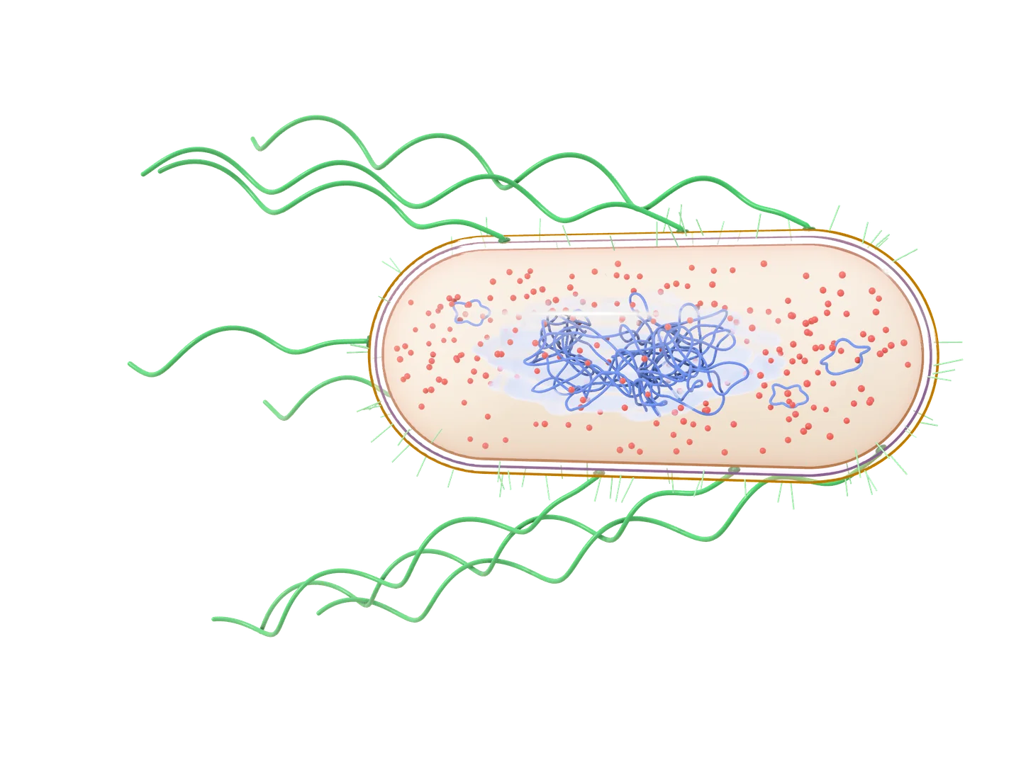
E. coli, a rod-shaped prokaryote, shown twice: as a model of the whole cell, which you can turn, and in a longitudinal section, a slice along its length. Its envelope has three layers: the outer membrane, a thin cell wall, and the plasma membrane. Inside, with no nucleus, a tangle of DNA, the nucleoid, fills the middle of the cytoplasm, with small rings of DNA, plasmids, and many ribosomes round it. Outside, short fimbriae cover the cell, and long wavy flagella rise all over it, each turned by a motor in the envelope, and stream back from it. Arrow keys turn it, plus and minus zoom, and Home brings back the first view.
Longitudinal section
Drag to turn it, pinch or scroll to zoom, and tap a part to read what it does.
E. coli, a prokaryote about 2 µm long, has no nucleus and no membrane-bound organelles: its one circular chromosome lies in the nucleoid, a region of the cytoplasm with no membrane round it. Its envelope is the plasma membrane, a thin wall of peptidoglycan and, outside that, the outer membrane of a gram-negative bacterium. Flagella, turned by motors in the envelope, drive it along, and fimbriae let it stick to surfaces and cells.
Simplified: not to scale: the envelope drawn thicker, and the flagella and fimbriae shorter than they are; the chromosome far shorter than it is: stretched out, it would be about 1.1 mm long, over 500 times the cell’s length; far fewer ribosomes than a real cell has; the flagella drawn apart, though while it swims they turn together in one bundle; in the model, the fimbriae shown only round its outline, where they stand out from it; the wall in carbohydrate’s colour, though its cross-links are peptides; the outer membrane drawn like the plasma membrane, though its outer layer holds lipopolysaccharide; ribosomes in RNA’s colour, though they are part protein.
10 sources
- Clark MA, Douglas M, Choi J. Biology 2e. OpenStax; 2018. 4.2 Prokaryotic Cells.
- Clark MA, Douglas M, Choi J. Biology 2e. OpenStax; 2018. 4.3 Eukaryotic Cells.
- Parker N, Schneegurt M, Thi Tu AH, et al. Microbiology. OpenStax; 2016. 3.3 Unique Characteristics of Prokaryotic Cells.
- Parker N, Schneegurt M, Thi Tu AH, et al. Microbiology. OpenStax; 2016. 3.4 Unique Characteristics of Eukaryotic Cells.
- Parker N, Schneegurt M, Thi Tu AH, et al. Microbiology. OpenStax; 2016. 14.3 Mechanisms of Antibacterial Drugs.
- Parker N, Schneegurt M, Thi Tu AH, et al. Microbiology. OpenStax; 2016. 9.2 Oxygen Requirements for Microbial Growth.
- Parker N, Schneegurt M, Thi Tu AH, et al. Microbiology. OpenStax; 2016. 15.2 How Pathogens Cause Disease.
- Clark MA, Douglas M, Choi J. Biology 2e. OpenStax; 2018. 7.4 Oxidative Phosphorylation.
- Clark MA, Douglas M, Choi J. Biology 2e. OpenStax; 2018. 14.2 DNA Structure and Sequencing.
- Escherichia coli. Wikipedia, citing Encyclopaedia Britannica and Kubitschek HE, J Bacteriol 1990.
Eukaryotes: an animal cell
Eukaryotic cells appeared about 2 billion years ago. They are larger, about 10 to 100 µm across against 0.1 to 5 µm for a prokaryote, and they divide their work among compartments bounded by membranes.
3D model
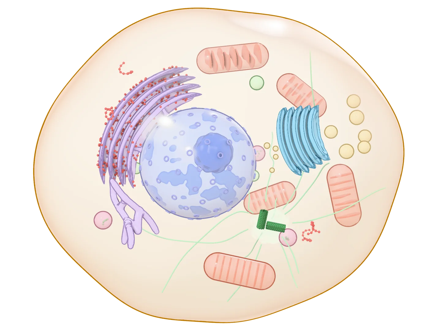
An animal cell, a eukaryote, shown twice: as a model of the whole cell, which you can turn, and in cross-section, as a slice through its middle. Its plasma membrane encloses the cytoplasm. The nucleus, inside a double envelope crossed by pores, holds the chromatin and a dense nucleolus. Beside it the rough endoplasmic reticulum lies in sheets studded with ribosomes, joined to the tubes of the smooth ER. The Golgi apparatus is a stack of curved sacs with vesicles round it, some carrying their load to the plasma membrane. Mitochondria show their folded inner membrane; lysosomes, peroxisomes and free ribosomes are scattered through the cytoplasm; and microtubules spread from the centrosome, with its two centrioles, by the nucleus. Arrow keys turn it, plus and minus zoom, and Home brings back the first view.
Cross-section
Drag to turn it, pinch or scroll to zoom, and tap a part to read what it does.
A eukaryotic cell, 10 to 100 µm across, keeps most of its DNA in a nucleus and shares its work among compartments bounded by membranes. Proteins for membranes and for export are made on the rough ER, modified in the Golgi apparatus and shipped in vesicles; mitochondria make most of the ATP, lysosomes digest, and microtubules hold the organelles in place and are the tracks they are carried along. Turn the model to see the cell whole; the cross-section shows what a slice through its middle cuts.
Simplified: not to scale: fewer, larger organelles than a real cell has, and fewer, far larger ribosomes; each organelle in a colour of its own, as textbooks paint them, though all their membranes are lipid; the nucleolus in DNA’s colour, though it holds RNA and protein too; in the cross-section, the rough ER and the Golgi show only the sacs the slice cuts, while the smooth ER and the microtubules are drawn whole; in the model, the nuclear envelope drawn as one layer, the chromatin only as the dense patches lining it, though it fills the nucleus, and the rough ER as a few nested sheets joined by short necks; of the cytoskeleton, only microtubules drawn; ribosomes in RNA’s colour, though they are part protein.
13 sources
- Silverthorn DU. Human Physiology, An Integrated Approach, 8th edition. Pearson; 2019. 3.3 Intracellular Compartments, 5.5 Vesicular Transport, and 22.4 Fed-State Metabolism.
- Clark MA, Douglas M, Choi J. Biology 2e. OpenStax; 2018. 4.2 Prokaryotic Cells.
- Clark MA, Douglas M, Choi J. Biology 2e. OpenStax; 2018. 4.3 Eukaryotic Cells.
- Clark MA, Douglas M, Choi J. Biology 2e. OpenStax; 2018. 4.4 The Endomembrane System and Proteins.
- Clark MA, Douglas M, Choi J. Biology 2e. OpenStax; 2018. 7.4 Oxidative Phosphorylation.
- Clark MA, Douglas M, Choi J. Biology 2e. OpenStax; 2018. 9.1 Signaling Molecules and Cellular Receptors.
- Clark MA, Douglas M, Choi J. Biology 2e. OpenStax; 2018. 10.2 The Cell Cycle.
- Clark MA, Douglas M, Choi J. Biology 2e. OpenStax; 2018. 23.1 Eukaryotic Origins.
- Parker N, Schneegurt M, Thi Tu AH, et al. Microbiology. OpenStax; 2016. 3.4 Unique Characteristics of Eukaryotic Cells.
- Parker N, Schneegurt M, Thi Tu AH, et al. Microbiology. OpenStax; 2016. 14.3 Mechanisms of Antibacterial Drugs.
- Betts JG, Young KA, Wise JA, et al. Anatomy and Physiology 2e. OpenStax; 2022. 3.2 The Cytoplasm and Cellular Organelles.
- Centrioles 2A. Digital Histology, an open educational resource.
- Tay-Sachs disease. MedlinePlus Genetics. National Library of Medicine (US).
- the nucleus, inside a double-membrane envelope pierced by pores, holds most of the cell's DNA, as linear chromosomes (46 in a human body cell);
- the rough endoplasmic reticulum, studded with ribosomes, makes membrane and secreted proteins;
- the smooth endoplasmic reticulum, with no ribosomes, makes lipids, among them the phospholipids of membranes and, in steroid-making cells, steroid hormones from cholesterol;
- the Golgi apparatus modifies proteins and lipids made in the ER and packages them into vesicles;
- mitochondria make most of the cell's ATP;
- lysosomes break down worn-out organelles and what the cell takes in.
| Prokaryotes | Eukaryotes | |
|---|---|---|
| Size | 0.1 to 5 µm | 10 to 100 µm |
| DNA | usually one circular chromosome, in the nucleoid | many linear chromosomes (46 in a human body cell), in a nucleus |
| Membrane-bound organelles | none | nucleus, ER, Golgi, mitochondria and others |
| Ribosomes | 70S | 80S (bacteria-like inside mitochondria) |
| Cell wall | in most bacteria, of peptidoglycan | none in animal cells; of cellulose in plants, of chitin in fungi |
| Examples | bacteria such as E. coli, and archaea | animals, plants, fungi, protists |
Where mitochondria came from
Mitochondria began as bacteria. An ancestral cell took in an alpha-proteobacterium, which lived on inside it and became the mitochondrion: endosymbiosis. Chloroplasts arose the same way, from cyanobacteria taken in by the ancestor of plants and of green and red algae.
Check yourselfAnswer, then say how sure you were: a lucky guess still counts as a weak point.
1Why is RNA, rather than DNA or protein, thought to have been life's first genetic molecule?
2Which of these do prokaryotic and eukaryotic cells both have?
3Where does Escherichia coli keep its chromosome?
4Which finding best supports the idea that mitochondria descend from bacteria?
5Why does penicillin harm bacteria but not the patient's own cells?
Linked topicsThe concepts this topic shares, and the topics they tie it to.
See on the mapBacterial cell wall
Prokaryotes such as E. coli, and the cell wall our cells lack.
- Antibiotics: classification; mechanisms, type and spectrum of action. Principles of the rational antibiotic therapy and prophylaxisPharmacologyAntibiotics are grouped by their target; the cell wall is one of the main ones.
- β-lactam antibiotics: overviewPharmacologyA β-lactam binds the penicillin-binding proteins, so new peptidoglycan isn’t cross-linked.
- PenicillinsPharmacologyThe first β-lactams, from benzylpenicillin to amoxicillin and piperacillin.
- CephalosporinsPharmacologyβ-lactams in generations: the first three reach ever more gram-negatives, the fifth MRSA too.
- Carbapenems and monobactams. GlycopeptidesPharmacologyCarbapenems are the broadest β-lactams; vancomycin blocks the wall by binding D-Ala-D-Ala itself.
- Bacterial community – acquired pneumoniaInternal medicineAmoxicillin treats most pneumococcal pneumonia.
- EndocarditisInternal medicineWeeks of intravenous β-lactams, or vancomycin, chosen by the organism.
- Hospital – acquired pneumonia and pneumonia in immunocompromised patientsInternal medicineCeftriaxone or ertapenem if resistance is unlikely; piperacillin–tazobactam or meropenem if it is likely.
Bacterial ribosome
Prokaryotes have 70S ribosomes, our cells 80S.
- Genetic code. TranslationBiopolymers: structure, synthesis and self-assemblyHow a ribosome reads mRNA into protein.
- TranslationBiochemistryNot written yetThe same process, step by step.
- Aminoglycosides (30S inhibitors)PharmacologyBind the 30S subunit and make it misread the code.
- Tetracyclines (30S inhibitors)PharmacologyBind the 30S subunit and stop new tRNAs docking.
- Macrolides and ketolides (50S inhibitors)PharmacologyBind the 50S subunit and block the growing chain.
- Lincosamides. Amphenicols (50S inhibitors)PharmacologyClindamycin and chloramphenicol bind the 50S subunit too.
- Bacterial community – acquired pneumoniaInternal medicineA macrolide or doxycycline covers atypical bacteria such as Mycoplasma.